Review
. 2007 Mar 15;23(4):303-10.
doi: 10.1016/s0828-282x(07)70760-2.

Echocardiographic evaluation of cardiac dyssynchrony

Affiliations
Review

Echocardiographic evaluation of cardiac dyssynchrony

Karim Serri et al. Can J Cardiol. .

Abstract

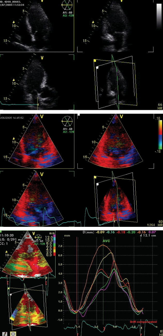
First described a decade ago, cardiac resynchronization therapy (CRT) has recently become a proven therapeutic strategy for refractory heart failure. Large clinical trials have shown a reduction in both morbidity and mortality in patients treated with CRT. Initial patient selection has relied mainly on electrocardiographic criteria, which allows identification of only 70% of responders. Accordingly, echocardiographic criteria were developed to identify mechanical dyssynchrony in an effort to improve patient selection. Multiple echocardiographic criteria have since been proposed, with no consensus as to which parameter better predicts CRT response. Although comparison studies using different criteria are underway, current evaluation of dyssynchrony should probably use an integrated multiparameter approach. The objective of the present article was to review the role of echocardiography in the evaluation of cardiac dyssynchrony in clinical practice.

Décrit pour la première fois il y a dix ans, le traitement de resynchronisation cardiaque (TRC) est récemment devenu une stratégie thérapeutique démontrée pour l’insuffisance cardiaque réfractaire. De grands essais cliniques ont révélé une réduction de la morbidité et de la mortalité chez les patients ayant subi un TRC. La sélection initiale des patients se fondait surtout sur des critères électrocardiographiques, par lesquels on ne repérait que 70 % des répondants. Par conséquent, les critères échocardiographiques étaient élaborés pour repérer la dyssynchronie mécanique afin d’améliorer la sélection des patients. De multiples critères échocardiographiques ont été proposés depuis, sans qu’on parvienne à un consensus sur les paramètres pour prévoir la réponse au TRC. Même si des études comparatives à l’aide d’autres critères sont en cours, une évaluation de la dyssynchronie devrait faire appel à une démarche multiparamétrique intégrée. Le présent article vise à analyser le rôle de l’échocardiographie dans l’évaluation de la dyssynchronie cardiaque en pratique clinique.


Figures

Figure 1)
Figure 1)
Measurement of aortic and pulmonary pre-ejection delays using conventional pulsed-wave Doppler. The systolic signals are obtained in a standard fashion: the pulsed-wave Doppler gate is placed in the left ventricular outflow tract just under the aortic valve from the apical five-chamber view for the aortic ejection signal, and in the right ventricular outflow tract just under the pulmonary valve from the parasternal short axis view for the pulmonary ejection signal. Measurements are performed by placing the first calliper at the QRS onset and the second calliper at the onset of the pulsed-wave Doppler ejection signal. A difference of more than 40 ms between right and left pre-ejection intervals confirms the presence of interventricular dyssynchrony
Figure 2)
Figure 2)
Comparison of the timings of posterior wall contraction with rapid filling phase. The M-mode recording is obtained from the parasternal long axis view, with the cursor at the tip of the mitral leaflets, and the delay (D1) from QRS onset to maximal posterior wall excursion is measured on the M-mode display. The transmitral pulsed-wave signal is obtained in a standard fashion, with the gate placed at the tip of the mitral leaflets in the apical four-chamber view and the delay (D2) from QRS onset to the mitral flow onset on the Doppler signal is measured. When contraction extends beyond the onset of rapid filling (D1 greater than D2), thus occurring during early diastole, intraventricular dyssynchrony is considered to be present
Figure 3)
Figure 3)
Measurement of septal-to-posterior wall motion delay between peak contraction of the interventricular septum and posterior wall on the M-mode display obtained from the parasternal long axis view, with the cursor at the tip of the mitral leaflets. Intraventricular dyssynchrony is present if the delay is more than 130 ms
Figure 4)
Figure 4)
Normal pulsed-wave tissue Doppler imaging recording obtained with the region of interest placed on the basal segment of the anterolateral wall from the apical four-chamber view. The Doppler tracing displays two systolic deflections, S wave and isovolumic contraction (IVC) signal, and two diastolic deflections, Ea and Aa waves, corresponding to early rapid filling and atrial contraction, respectively
Figure 5)
Figure 5)
Measurement of the electromechanical delay (EMD) and electrosystolic delay (ESD) using pulsed-wave tissue Doppler imaging with sampling in the basal segments of opposite left ventricular walls (the interventricular septum and the anterolateral wall) in the apical four-chamber view. The EMD is measured from the QRS onset on the electrocardiogram tracing to the onset of the S wave, and the ESD is measured from the QRS onset to the peak of the S wave. A difference of more than 40 ms between opposite walls for either EMD or ESD indicates intraventricular dyssynchrony
Figure 6)
Figure 6)
Numerical tissue Doppler imaging tracings reconstructed from colour tissue Doppler imaging acquisitions from the apical four-chamber view. This method allows direct, simultaneous comparison of systolic velocities and timing between opposite left ventricular walls, in this case the basal septum (yellow) and basal lateral (green) wall
Figure 7)
Figure 7)
Myocardial deformation, or strain tracings, showing simultaneous longitudinal deformation of basal lateral (green) and basal septal (yellow) left ventricular walls from the apical four-chamber view. Intraventricular dyssynchrony is present, as evidenced by one of the segments contracting after aortic valve closure. DLC Delayed longitudinal contraction
Figure 8)
Figure 8)
Apical four-chamber view using the tissue synchronization imaging mode. This parametric imaging modality allows direct visualization of segments with early displacement (in green) and late activation (in yellow-orange)
Figure 9)
Figure 9)
Colour map of intraventricular dyssynchrony derived from real-time three-dimensional acquisitions before and after cardiac resynchronization therapy. Early activated regions are represented in blue, while late activated regions are coloured red. Left panel: spontaneous rhythm; Right panel: Biventricular pacing ‘on’
Figure 10)
Figure 10)
Multislice imaging with simultaneous acquisition of three standard apical views (two-, three- and four-chamber). This technique allows direct comparison of numeric tissue Doppler imaging tissue tracking tracings for all 12 basal and mid-left ventricular segments from the apical window (shown on right, simultaneous tissue tracking tracings of the six basal left ventricular segments). AVC Aortic valve closure
Figure 11)
Figure 11)
Diagram comparing the different phases of systole assessed by each dyssynchrony parameter. Electromechanical delay (EMD) and electrosystolic delay (ESD) evaluate early- and mid-systole, respectively, whereas delayed longitudinal contraction (DLC) assesses end-systole. ECG Electrocardiogram

References

    1. Hunt SA, Baker DW, Chin MH, et al. American College of Cardiology/American Heart Association Task Force on Practice Guidelines (Committee to Revise the 1995 Guidelines for the Evaluation and Management of Heart Failure); International Society for Heart and Lung Transplantation; Heart Failure Society of America ACC/AHA guidelines for the evaluation and management of chronic heart failure in the adult: Executive summary: A report of the American College of Cardiology/American Heart Association Task Force on Practice Guidelines (Committee to Revise the 1995 Guidelines for the Evaluation and Management of Heart Failure): Developed in collaboration with the International Society for Heart and Lung Transplantation; endorsed by the Heart Failure Society of America. Circulation. 2001;104:2996–3007. - PubMed
    1. Grines CL, Bashore TM, Boudoulas H, Olson S, Shafer P, Wooley CF. Functional abnormalities in isolated left bundle branch block. The effect of interventricular asynchrony. Circulation. 1989;79:845–53. - PubMed
    1. Cazeau S, Ritter P, Bakdach S, Lazarus A, et al. Four chamber pacing in dilated cardiomyopathy. Pacing Clin Electrophysiol. 1994;17:1974–9. - PubMed
    1. Gregoratos G, Abrams J, Epstein AE, et al. American College of Cardiology/American Heart Association Task Force on Practice Guidelines/North American Society for Pacing and Electrophysiology Committee to Update the 1998 Pacemaker Guidelines ACC/AHA/NASPE 2002 guideline update for implantation of cardiac pacemakers and antiarrhythmia devices: Summary article: A report of the American College of Cardiology/American Heart Association Task Force on Practice Guidelines (ACC/AHA/NASPE Committee to Update the 1998 Pacemaker Guidelines) Circulation. 2002;106:2145–61. - PubMed
    1. Auricchio A, Stellbrink C, Sack S, et al. Pacing Therapies in Congestive Heart Failure (PATH-CHF) Study Group Long-term clinical effect of hemodynamically optimized cardiac resynchronization therapy in patients with heart failure and ventricular conduction delay. J Am Coll Cardiol. 2002;39:2026–33. - PubMed

MeSH terms