Review
. 2007 Mar;64(6):692-703.
doi: 10.1007/s00018-007-6522-3.

Epicardium-derived cells in cardiogenesis and cardiac regeneration

Affiliations
Review

Epicardium-derived cells in cardiogenesis and cardiac regeneration

E M Winter et al. Cell Mol Life Sci. 2007 Mar.

Abstract

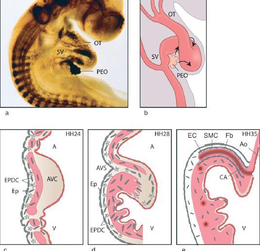
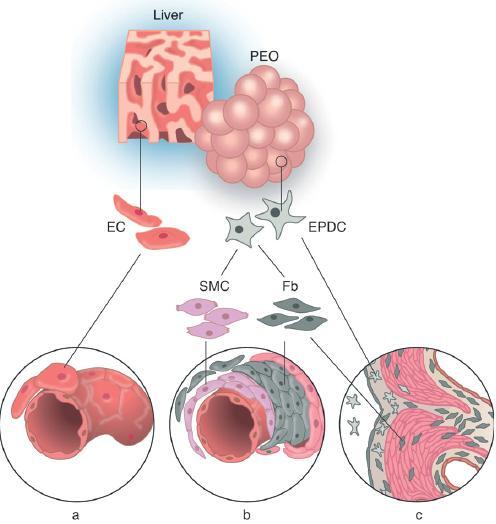
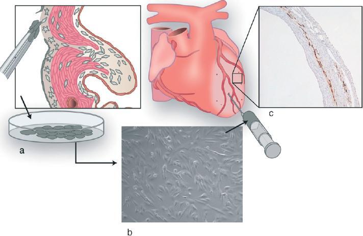
During cardiogenesis, the epicardium grows from the proepicardial organ to form the outermost layer of the early heart. Part of the epicardium undergoes epithelial-mesenchymal transformation, and migrates into the myocardium. These epicardium-derived cells differentiate into interstitial fibroblasts, coronary smooth muscle cells, and perivascular fibroblasts. Moreover, epicardium-derived cells are important regulators of formation of the compact myocardium, the coronary vasculature, and the Purkinje fiber network, thus being essential for proper cardiac development. The fibrous structures of the heart such as the fibrous heart skeleton and the semilunar and atrioventricular valves also depend on a contribution of these cells during development. We hypothesise that the essential properties of epicardium-derived cells can be recapitulated in adult diseased myocardium. These cells can therefore be considered as a novel source of adult stem cells useful in clinical cardiac regeneration therapy.


Figures

Figure 1
Figure 1
Spreading and migration of EPDCs. (a) Whole-mount quail embryo (HH16) stained for HNK1, showing a clearly demarcated proepicardial organ (PEO) at the venous pole of the heart, (b) Schematic representation indicating (arrows) the direction of growth of the epicardium over the myocardial tube. (c–e) Schematic drawing of increasing ages with the migration pattern of the EPDCs. (c) HH24: epicardial cells cover the heart tube, and EPDCs (star shaped, grey) enter the myocardium and, through gaps, the subendocardial layer. The endocardial cushion is still devoid of EPDCs. (d) HH28: the compact myocardium is formed, and EPDCs have entered all cardiac components. Note the contribution to the formed atrioventricular sulcus and the endocardial cushions, (e) HH35: the coronary vasculature has grown into the aorta, and EPDCs through EMT (cuboid cells) now also contribute to the coronary arterial vascular wall. OT, outflow tract; SV, sinus venosus; PEO, proepicardial organ; AVC, atrioventricular cushion; EPDC, epicardium-derived cell; Ep, epicardium; V, ventricle; A, atrium; AVS, atrioventricular sulcus; EC, endothelial cell; SMC, smooth muscle cell; Fb, fibroblast, CA, coronary artery; Ao, aorta.
Figure 2
Figure 2
Differentiation of EPDCs. (a) The coronary endothelial cells (EC, pink) are derived from liver sinusoidal cells and grow into the myocardium along with the EPDCs. (b) The epicardium is derived from the proepicardial organ (PEO), and EPDCs are formed through epithelial mesenchymal transformation. The EPDCs (star shaped, grey) are still undifferentiated and have the potential to form smooth muscle cells (SMC, purple) and fibroblasts (Fb, grey). These cells form the media and adventitia of the coronary vessels, (c) The fibroblasts form the interstitial cells of the myoardium as well as the cells of the fibrous skeleton of the heart. Their role in valve differentiation is unknown. PEO, proepicardial organ; EC, endothelial cell, EPDC, epicardium-derived cell; SMC, smooth muscle cell; Fb, fibroblast.
Figure 3
Figure 3
Illustration of EPDC transplantation experiments, (a) Adult epicardial cells are cultured, (b) Cultured epicardial cells are injected into the ischemic area and border zone of the left ventricular wall, (c) Histological section (10×) of the ischemic left ventricular wall after immunohistochemical staining against enhanced green fluorescent protein (eGFP), showing injected eGFP-transduced EPDCs.

References

    1. DeRuiter M. C., Poelmann R. E., VanderPlas-de Vries I., Mentink M. M. T., Gittenberger-de Groot A. C. The development of the myocardium and endocardium in mouse embryos. Fusion of two heart tubes? 1992;185:461–473. - PubMed
    1. Gittenberger-de Groot A. C., Bartelings M. M., DeRuiter M. C., Poelmann R. E. Basics of cardiac development for the understanding of congenital heart malformations. Pediatr. Res. 2005;57:169–176. - PubMed
    1. Kurkiewicz, T. (1909) [Zur Histogenese des Hersmuzkels der Wirbeltiere]. Bull. Int. Acad. Sci. Cracovie 148–191.
    1. Kölliker A. Entwicklungsgeschichte des Menschen und der Thiere. Leipzig: Engelmann; 1879.
    1. De Haan R. L. In: Organogenesis. De Haan R. L., Ursprung H., editors. New York: Holt, Rinehart and Winston; 1965.

LinkOut - more resources