Review
. 2007 Mar;64(6):704-18.
doi: 10.1007/s00018-007-6523-2.

Regulation of cardiomyocyte differentiation of embryonic stem cells by extracellular signalling

Affiliations
Review

Regulation of cardiomyocyte differentiation of embryonic stem cells by extracellular signalling

A A Filipczyk et al. Cell Mol Life Sci. 2007 Mar.

Abstract

Investigating the signalling pathways that regulate heart development is essential if stem cells are to become an effective source of cardiomyocytes that can be used for studying cardiac physiology and pharmacology and eventually developing cell-based therapies for heart repair. Here, we briefly describe current understanding of heart development in vertebrates and review the signalling pathways thought to be involved in cardiomyogenesis in multiple species. We discuss how this might be applied to stem cells currently thought to have cardiomyogenic potential by considering the factors relevant for each differentiation step from the undifferentiated cell to nascent mesoderm, cardiac progenitors and finally a fully determined cardiomyocyte. We focus particularly on how this is being applied to human embryonic stem cells and provide recent examples from both our own work and that of others.


Figures

Figure 1
Figure 1
A model of embryonic stem cell differentiation towards the cardiomyocyte lineage based on current mouse ESC differentiation methods. Pluripotent ESCs are maintained through transcription factors Oct-4, Nanog and Sox-2. Given appropriate signals from EB culture or a 2D culture system, the cells differentiate into a primitive, pre-hemangioblast type cell with broad mesodermal potential (blue arrow). These cells are characterised by the expression of key transcription factors like brachyury and MESP and may still retain the expression of pluripotency associated genes. From these cells, a Flk-1-positive subpopulation appears with hemangiblast-like differentiation activity highlighted by a robust ability to form blood cells (large orange arrow). However, these cells still retain remarkable plasticity and can give rise to increasingly more restricted Flk-1-positive progenitors (blue arrow). More restricted Flk-1-positive cells may predominantly differentiate into endothelial cells (large orange arrow) and less readily (small orange arrow) to cardiac cells. Immunosorting or magnetic cell separation for Flk-1 and other surface receptors like CXCR4 enables the enrichment of a more defined progenitor cell subpopulation which differentiates predominantly into beating cardiomyocytes (large orange arrow) with a limited capacity to form endothelial cells (small orange arrow).
Figure 2
Figure 2
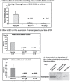
Pharmacological GSK-3 inhibitor BIO activates Wnt signalling and blocks cardiac differentiation of HESCs. To induce cardiac differentiation, HES2 cells were co-cultured with END2 feeders [35] and cultured in a serum-free medium for at least 12 days [108]. At day 5, the medium was changed, and BIO was added at a concentration of 1 or 2 µM in serum-free medium (or serum-free medium only, for control). At day 9 and 12, the co-cultures were refreshed with serum-free medium only, then harvested for RNA or protein isolation. (a) Scoring of the number of areas of beating muscle in the co-cultures at day 12 showed that BIO was inhibitory. On average, the reduction in the number of beating areas was 18-fold in the presence of 1 µM BIO and 61-fold in the presence of 2 µM BIO, respectively, compared with control values. (b) Determining the expression of two cardiac proteins, α-actinin and βMHC, by real-time polymerase chain reaction (PCR) showed a reduction in the presence of BIO addition by 10- and 11- fold, respectively, in the presence of 1 µM BIO and 164- and 93-fold, respectively, in the presence of 2 µM BIO. The cycle number at which the reaction crossed an arbitrary threshold (Ct) was determined for each gene. The relative number of messenger RNA levels was determined by 2-DCt. Relative gene expression was normalized to hARP expression. (c) Analysis of the effect of BIO on cardiac differentiation in 17-day co-cultures by Western blotting showed a strong decrease in the expression of the cardiac marker tropomyosin in the presence of BIO. p values were determined using the Man-Whitney test.

References

    1. Beltrami A.P., Torella D., Baker M., Limana F., Chimenti S., Kasahara H., Rota M., Musso E., Urbanek K., Leri A., Kajstura J., et al. Adult cardiac stem cells are multipotent and support myocardial regeneration. Cell. 2003;114:763–776. - PubMed
    1. Urbanek K., Cesselli D., Rota M., Nascimbene A., De Angelis A., Hosoda T., Bearzi C., Boni A., Bolli R., Kajstura J., et al. Stem cell niches in the adult mouse heart. Poc. Natl. Acad. Sci. USA. 2006;103:9226–9231. - PMC - PubMed
    1. Laugwitz K. L., Moretti A., Lam J., Gruber P., Chen Y., Woodard S., Lin L. Z., Cai C. L., Lu M. M., Reth M., Platoshyn O., Yuan J. X., Evans S., Chien K. R. Postnatal isl1+ cardioblasts enter fully differentiated cardiomyocyte lineages. Nature. 2005;433:647–653. - PMC - PubMed
    1. Passier R., Mummery C. Origin and use of embryonic and adult stem cells in differentiation and tissue repair. Cardiovasc. Res. 2003;58:324–335. - PubMed
    1. Passier R., Mummery C. Cardiomyocyte differentiation from embryonic and adult stem cells. Curr. Opin. Biotechnol. 2005;16:498–502. - PubMed

Publication types

LinkOut - more resources