

Review
doi: 10.1101/sqb.2006.71.053.
Structural biology of RNA silencing and its functional implications
Affiliations
- PMID: 17381284
- PMCID: PMC4689314
- DOI: 10.1101/sqb.2006.71.053
Item in Clipboard
Review
Structural biology of RNA silencing and its functional implications
Cold Spring Harb Symp Quant Biol.
2006.
Abstract
We outline structure-function contributions from our laboratories on protein-RNA recognition events that monitor siRNA length, 5 -phosphate and 2-nucleotide 3 overhangs, as well as the architecture of Argonaute, its externally bound siRNA complex, and Argonaute-based models involving guide-strand-mediated mRNA binding, cleavage, and release.
Figures
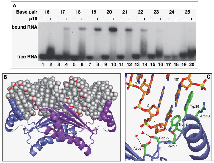
(A) Electrophoretic mobility-shift assays defining the length dependence of self-complementary siRNA duplexes for complex formation with viral suppressor p19. (B) The crystal structure of the TBSV p19–21-mer siRNA duplex (19-bp and 2-nucleotide 3′-overhang) (Ye et al. 2003). The RNA is shown in a space-filling representation with phosphorus atoms in red. The 2-nucleotide 3′-overhang bases are disordered and not shown. The protein is shown in a ribbon representation except for the pair of bracketing Trp residues at either end, which are shown in a space-filling representation. (C) Stick (RNA) and ribbon (protein) representation of stacking and hydrogen-bonding recognition of one RNA end in the complex. (Reprinted, with permission, from Ye et al. 2003 [Nature Publishing Group].)
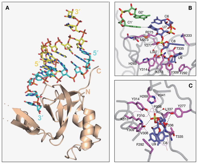
(A) The structure of one PAZ domain interacting with an siRNA-like end in the crystal structure of the human Ago1 PAZ-9-mer siRNA duplex (7-bp and 2-nucleotide 3′-overhang) (Ma et al. 2004). The complex is shown in stick (RNA) and ribbon (protein) representation. The RNA strand bound by its 3′-end is colored blue, whereas the strand bound by the 5′-end is colored yellow. (B) A stick representation of the interaction between the PAZ domain with the duplex terminus and 3′-overhang segments. Note the change in the RNA phosphodiester backbone between duplex and overhang segments. (C) Interactions between the 2-nucleotide 3′-overhang of the RNA and the walls of the conserved binding pocket on the PAZ domain. The residues directly contacting the RNA are shown in a stick representation, and other residues contributing to the hydrophobic pocket are shown as thin lines. Key hydrogen-bonding interactions from protein side chains to the internucleotide phosphate and the sugar hydroxyls are highlighted by dashed lines. (Reprinted, with permission, from Ma et al. 2004 [Nature Publishing Group].)
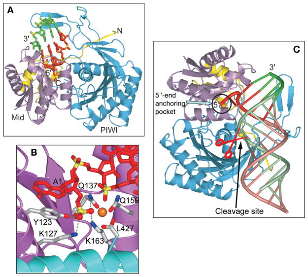
(A) The crystal structure of the A. fulgidus Piwi-21-mer siRNA duplex (19-bp and 2-nucleotide 3′-overhang) (Ma et al. 2005). The Piwi protein, shown in a ribbon representation, consists of a yellow-colored aminoterminal element (1–37), a magenta-colored Mid domain (38–167), and a cyan-colored PIWI domain (168–427). The segment of bound RNA that can be monitored in the structure is shown in a stick representation, with the protein interacting strand containing the 5′-phosphate colored red and its complementary partner strand colored green. (B) The 5′-phosphate-binding site in the complex. The 5′-phosphate is positioned in a basic pocket lined by K127, K163, Q137, and Q159 of the Mid domain and the carboxyterminal carboxylate from the PIWI domain, and a bound divalent cation in orange. Bases A1 and G2 are splayed apart, with unpaired A1 stacked on Y123. (C) Model of the A. fulgidus Piwi protein bound to a 5′-phosphate-containing siRNA duplex complex. The 5′-end nucleotide and 4-bp duplex (colored in red for the guide strand and green for the target strand) observed in the crystal structure of the complex were extended by an A-form duplex (colored tan for the guide strand and light green for the target strand). The 5′-end anchoring pocket and putative catalytic site are circled and labeled by an arrow, respectively, and the phosphate at the cleavage site is marked by a yellow ball. (Reprinted, with permission, from Ma et al. 2005 [Nature Publishing Group].)
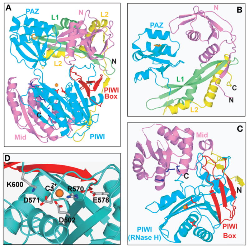
The crystal structure of A. aeolicus Argonaute (Yuan et al. 2005). The protein was crystallized in the presence of rU8, but the RNA was disordered and could not be traced. The view emphasizes the bilobal topology of Ago, with the aminoterminal PAZ-containing lobe on top and the carboxyterminal PIWI-containing lobe on the bottom. The various domains and linkers are numbered and color-coded. (B) Relative arrangements of the magenta-colored N (1–108), green-colored linker L1 (108–165), cyan-colored PAZ (166–262), and segment of yellow-colored linker L2 (263–311) within the PAZ-containing lobe. A Trp ring lining the PAZ-binding pocket is colored orange. (C) Relative arrangements of segment of the yellow-colored linker L2 (312–334), magenta-colored Mid domain (335–488), and cyan-colored PIWI domain (489–706) within the PIWI-containing lobe. Lys residues lining the 5′-phosphate- binding pocket on the Mid domain are colored dark blue. The DDE triad residues lining the catalytic binding pocket on the PIWI domain are colored red. The PIWI box (622–650) is colored red. (D) Relative positioning of invariant catalytic acidic D502, D571, and E578 residues and bound Ca cation on the surface of the RNase H fold of the PIWI domain. Invariant acidic R570 is also positioned in the catalytic pocket, whereas conserved basic K600 is directed toward the catalytic pocket. The Ca cation is also coordinated by D683, which is a His residue in hAgo2. (Reprinted, with permission, from Yuan et al. 2005 [© Elsevier].)
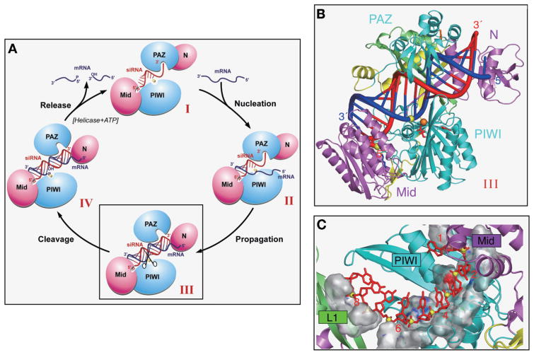
(A) Schematic of the catalytic cycle involving guide-RNA-dictated mRNA loading, cleavage, and product release within the context of the Ago scaffold (Yuan et al. 2005). The states represented by conformers I, II, III, and IV are described in the text. The mRNA nucleation step corresponds to the transition from conformer I to II, the mRNA propagation step corresponds to the transition from conformer II to III, the mRNA cleavage step corresponds to the transition from conformer III to IV, and the product release step corresponds to the transition from conformer IV back to conformer I. (B) A view of the model of the AaAgo–DNA/RNA hybrid complex. The orientation of Ago is rotated along the vertical axis relative to the one shown in Fig. 4A. The color coding and labeling of domains and linkers, as well as key amino acids, are the same as listed in Fig. 4A. The hybrid duplex between the guide DNA strand (colored red) and the mRNA strand (colored blue) is shown in a tubular representation, with a thicker diameter for the sugar-phosphate backbone and thinner diameter for the bases. The cleavable phosphate positioned between residues 10 and 11 on the mRNA strand (as counted from the 5′-end of the guide strand) is shown by a yellow ball. (C) The phosphodiester backbone corresponding to positions 2–8 from the 5′-end of the guide strand are positioned within a trough-like segment of the Mid and PIWI domains in the model of the complex. The guide strand is shown in red, with phosphorus atoms as yellow balls. The trough is shown in a surface representation and exhibits surface complementarity with the sugar-phosphate backbone of the 5′-end region of the guide strand. (Reprinted, with permission, from Yuan et al. 2005 [© Elsevier].)
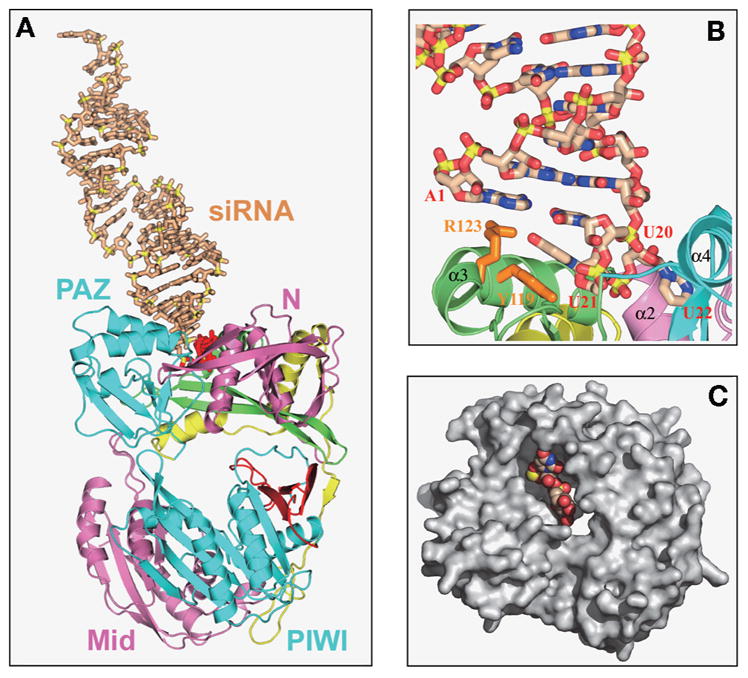
(A) The crystal structure of A. aeolicus Ago bound externally to a 22-mer siRNA (20-bp and 2-nucleotide 3′-overhang) (Yuan et al. 2006). The Ago protein is shown in a ribbon representation with the color coding and labeling of domains and linkers the same as listed in Fig. 4A. The externally bound siRNA, shown in a stick representation, is shown in beige with backbone phosphorus atoms in yellow, except for the 2-nucleotide overhang at the bound end, which is colored red. (B) A view of interactions between the 2-nucleotide overhang at one end of the externally bound 22-mer siRNA and the cavity positioned on the outer surface of the PAZ-containing lobe of AaAgo. The overhang base U21 stacks on the aromatic ring of orange-colored Y119, whereas the overhang base U22 is inserted into a cavity whose walls involve segments of N domain, linker L1, and the PAZ domain. (C) Surface representation highlighting the cavity located on the outward-pointing face of the PAZ-containing lobe of the siRNA-bound AaAgo. The bound 2-nucleotide 3′-overhang is shown in a space-filling representation. (Reprinted, with permission, from Yuan et al. 2006 [© Elsevier].)
Similar articles
-
The Argonautes.Cold Spring Harb Symp Quant Biol. 2006;71:67-72. doi: 10.1101/sqb.2006.71.048. Cold Spring Harb Symp Quant Biol. 2006. PMID: 17381282 Review.
-
Molecular biology. Argonaute journeys into the heart of RISC.Science. 2004 Sep 3;305(5689):1409-10. doi: 10.1126/science.1103076. Science. 2004. PMID: 15353786 No abstract available.
-
Structural basis for 5'-end-specific recognition of guide RNA by the A. fulgidus Piwi protein.Nature. 2005 Mar 31;434(7033):666-70. doi: 10.1038/nature03514. Nature. 2005. PMID: 15800629 Free PMC article.
-
Structural insights into mRNA recognition from a PIWI domain-siRNA guide complex.Nature. 2005 Mar 31;434(7033):663-6. doi: 10.1038/nature03462. Nature. 2005. PMID: 15800628 Free PMC article.
-
Argonaute proteins: mediators of RNA silencing.Mol Cell. 2007 Jun 8;26(5):611-23. doi: 10.1016/j.molcel.2007.05.001. Mol Cell. 2007. PMID: 17560368 Review.
Cited by
-
Knocking down Insulin Receptor in Pancreatic Beta Cell lines with Lentiviral-Small Hairpin RNA Reduces Glucose-Stimulated Insulin Secretion via Decreasing the Gene Expression of Insulin, GLUT2 and Pdx1.Int J Mol Sci. 2018 Mar 26;19(4):985. doi: 10.3390/ijms19040985. Int J Mol Sci. 2018. PMID: 29587416 Free PMC article.
-
Structure of an argonaute silencing complex with a seed-containing guide DNA and target RNA duplex.Nature. 2008 Dec 18;456(7224):921-6. doi: 10.1038/nature07666. Nature. 2008. PMID: 19092929 Free PMC article.
-
Nucleation, propagation and cleavage of target RNAs in Ago silencing complexes.Nature. 2009 Oct 8;461(7265):754-61. doi: 10.1038/nature08434. Nature. 2009. PMID: 19812667 Free PMC article.
-
Structural basis for RNA-silencing suppression by Tomato aspermy virus protein 2b.EMBO Rep. 2008 Aug;9(8):754-60. doi: 10.1038/embor.2008.118. Epub 2008 Jul 4. EMBO Rep. 2008. PMID: 18600235 Free PMC article.
-
A multifunctional human Argonaute2-specific monoclonal antibody.RNA. 2008 Jun;14(6):1244-53. doi: 10.1261/rna.973808. Epub 2008 Apr 22. RNA. 2008. PMID: 18430891 Free PMC article.
References
-
- Aravin AA, Lagos-Quintana M, Yalcin A, Zavolan M, Marks D, Snyder B, Gaasterland T, Meyer J, Tuschl T. The small RNA profile during Drosophila melanogaster development. Dev Cell. 2003;5:337. - PubMed
-
- Aravin A, Gaidatzis D, Pfeffer S, Lagos-Quintana M, Ladgraf P, Iovino N, Morris P, Brownstein MJ, Kuramochi-Miyagawa S, Nakano T, et al. A novel class of small RNAs bind to MLL1 protein in mouse testis. Nature. 2006;442:203. - PubMed
-
- Bartel DP. MicroRNAs: Genomics, biogenesis, mechanism and function. Cell. 2004;116:281. - PubMed
-
- Baulcombe D. RNA silencing in plants. Nature. 2004;431:356. - PubMed
-
- Bernstein E, Caudy AA, Hammond SM, Hannon GJ. Role for a bidentate ribonuclease in the initiation step of RNA interference. Nature. 2001;409:363. - PubMed
Publication types
MeSH terms
Substances
Grants and funding
LinkOut - more resources
Full Text Sources
Other Literature Sources