

Review
doi: 10.1021/jm00081a001.
Adenosine receptors: pharmacology, structure-activity relationships, and therapeutic potential
Affiliations
- PMID: 1738138
- PMCID: PMC3476067
- DOI: 10.1021/jm00081a001
Item in Clipboard
Review
Adenosine receptors: pharmacology, structure-activity relationships, and therapeutic potential
J Med Chem.
.
No abstract available
Figures
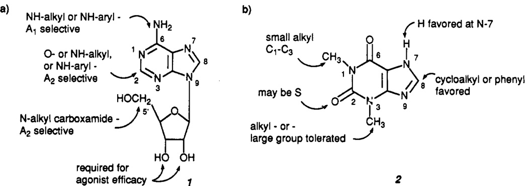
The structures of adenosine (1) and theophylline (2), showing the effects of structural modifications at various sites on receptor binding.

Computer-generated models of the N6 regions of adenosine A1 and A2 receptors. For the A1 model, areas are indicated where hydrophobic substituents may lead to enhanced affinity. These are designated S1, S1A, S2, S3, S3A, A(ryl), B(ulk), and C(ycloalkyl). In both models, shaded areas indicate the receptor boundaries (adapted from ref and 31).
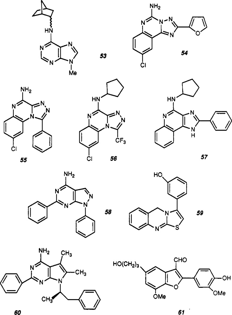
The structures of non-xanthine adenosine antagonists. (See text and Table III for description.)

Computer-generated model of the antagonist binding site of the adenosine A1 receptor. Indicated is the molecular electrostatic potential (at the +5 and −5 kcal/mol level) in the plane of the 6:5-fused heterocycle that is common to xanthines and many non-xanthine adenosine antagonists. R (corresponds to the N6 position of adenosine) and dotted lines indicate regions where hydrophobic substitution may enhance affinity. N7 is thought to act as a hydrogen bond acceptor (adapted from ref 49).
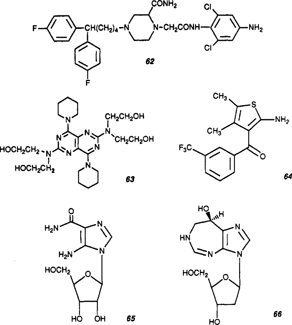
Agents for indirect modulation of adenosine function through transport (62 and 63) or metabolic processes (65 and 66), or at an allosteric site on the A1 receptor (64). See text for description.

Structure of Ap4A.

A proposed model (121) for adenosine receptors deduced from the primary sequences, showing features common to both A1 and A2 receptors, including the seven transmembrane helices typical of G-protein-coupled receptors (I–VII), three extracellular loops (EI–III), three cytoplasmic loops (CI–III), two histidinyl residues (H) (possibly involved in ligand binding) a sodium binding site (Na), and sites for phosphorylation of serine and threonine residues (P) and glycosylation. Phosphorylation sites on the C-terminus apply to the A2-sequence only. A site for acylation (Ac) applies to A1 receptors only. The C-terminal sequences (beyond H-7) are approximately 34 and 119 residues in length for canine A1 and A2 receptors, respectively.
References
-
- Daly JW. Adenosine receptors: targets for future drugs. J. Med. Chem. 1982;25:197–207. - PubMed
-
- Williams M. Adenosine: the prototypic neuromodulator. Neurochem. Intern. 1989;14:249–264. - PubMed
-
- Maenhaut C, van Sande SJ, Libert F, Abramowicz M, Parmentier M, Vanderhaegen JJ, Dumont JE, Vassart G, Schiffmann S. RDC8 codes for an adenosine A2 receptor with physiological constitutive activity. Biochem. Biophys. Res. Commun. 1990;173:1169–1178. - PubMed
-
- Hollenberg MD. Receptor triggering and receptor regulation: Structure–activity relationship from the receptor’s point of view. J. Med. Chem. 1990;33:1275–1281. - PubMed
Publication types
MeSH terms
Substances
Grants and funding
LinkOut - more resources
Full Text Sources
Other Literature Sources
Chemical Information