

Differentiation of cardiomyocytes requires functional serine residues within the amino-terminal domain of desmin
- PMID: 17381546
- PMCID: PMC7615843
- DOI: 10.1111/j.1432-0436.2007.00163.x
Differentiation of cardiomyocytes requires functional serine residues within the amino-terminal domain of desmin
Abstract
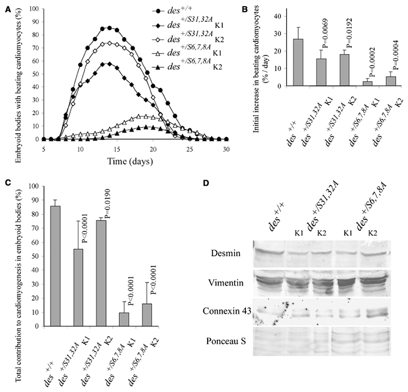
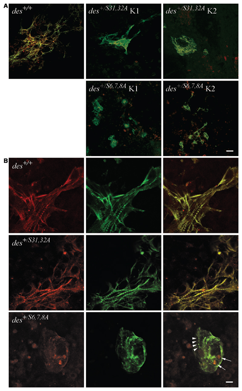
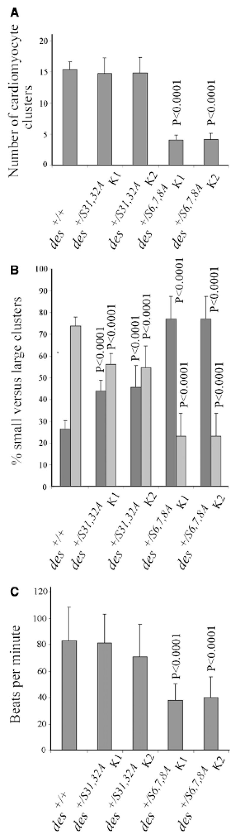
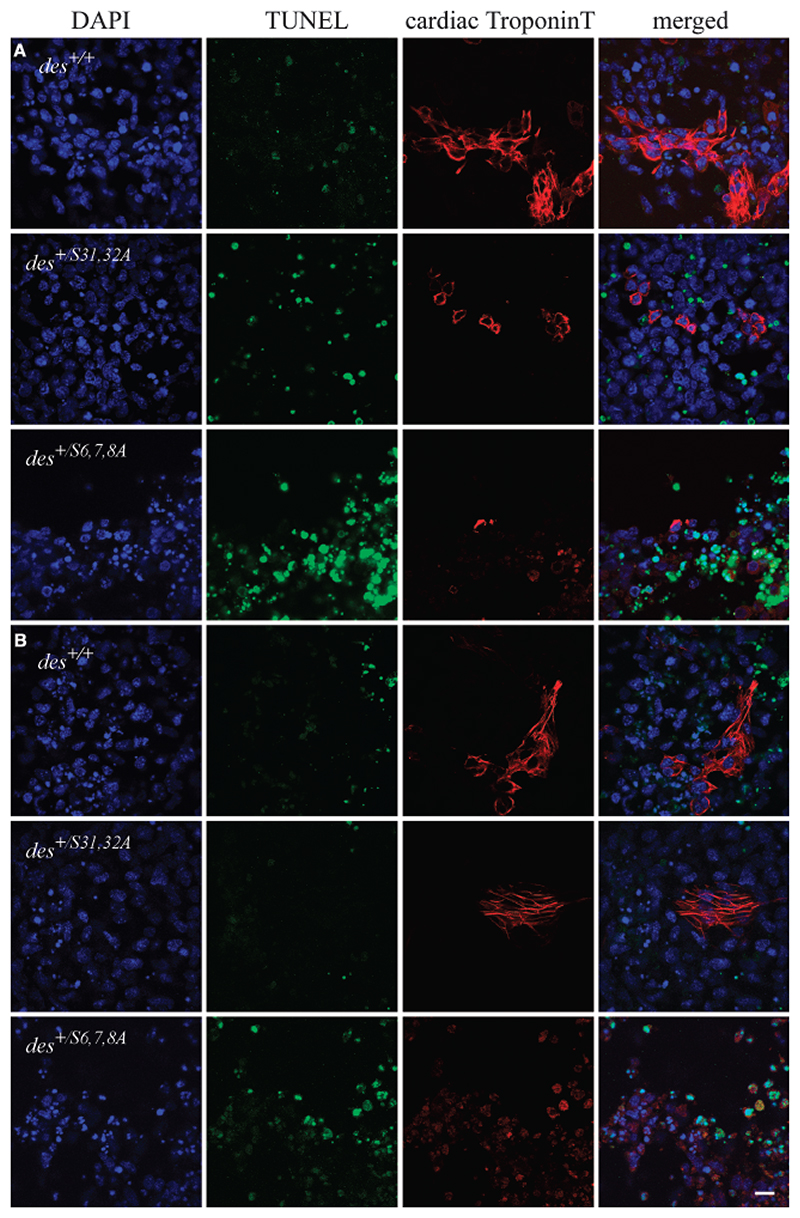
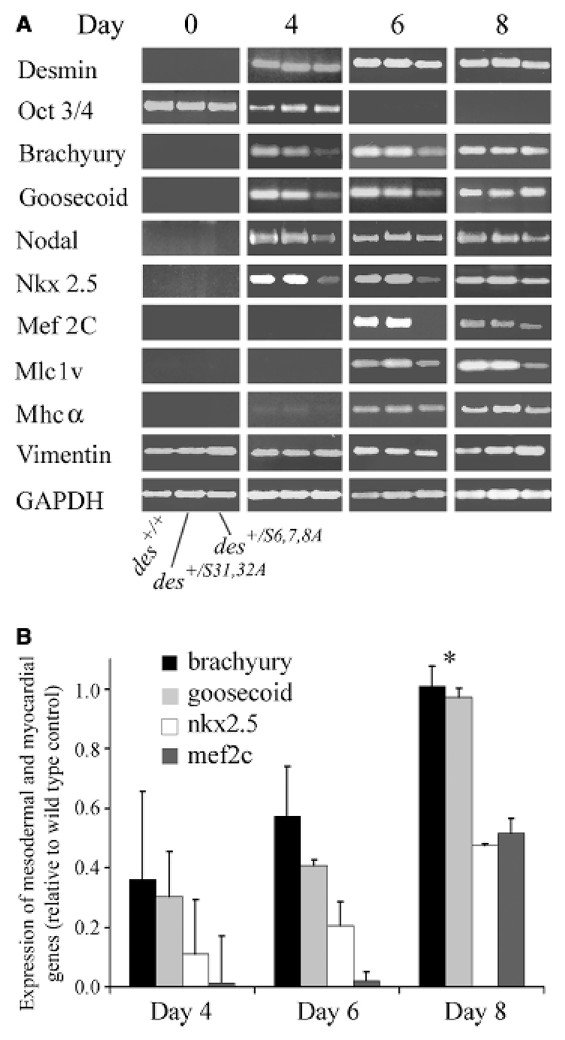
Desmin contributes to the stability of the myocardium and its amino-terminal domain influences intermediate filament formation and interacts with a variety of proteins and DNAs. Specific serine residues located in this domain are reversibly phosphorylated in a cell cycle and developmental stage-dependent manner as has been demonstrated also for other cytoplasmic type III intermediate filament proteins. Although absence of desmin apparently does not affect cardiomyogenesis, homozygous deletion of the amino-terminal domain of desmin severely inhibited in vitro cardiomyogenesis. To demonstrate the significance of phosphorylation of this domain in cardiomyogenic commitment and differentiation, we inhibited phosphorylation of serine residues 6, 7, and 8 by mutation to alanine, and investigated early cardiomyogenesis in heterozygous embryoid bodies. As control, serine residues 31 and 32, which are not phosphorylated by kinases mutating serine residues 6, 7, and 8, were mutated to alanine in a second set. Desmin(S6,7,8A) interfered with cardiomyogenesis and myofibrillogenesis in a dominant negative fashion, whereas desmin(S31,32A) produced only a mild phenotype. Desmin(S6,7,8A) led to the down-regulation of the transcription factor genes brachyury, goosecoid, nkx2.5, and mef2C and increased apoptosis of presumptive mesoderm and differentiating cardiomyocytes. Surviving cardiomyocytes which were few in number had no myofibrils. Demonstration that some but not any mutant desmin interfered with the very beginning of cardiomyogenesis suggests an important function of temporarily phosphorylated serine residues 6, 7, and 8 in the amino-terminal domain of desmin in cardiomyogenic commitment and differentiation.
Figures
Similar articles
-
Desmin stimulates differentiation of cardiomyocytes and up-regulation of brachyury and nkx2.5.Differentiation. 2007 Sep;75(7):605-15. doi: 10.1111/j.1432-0436.2007.00162.x. Epub 2007 Mar 23. Differentiation. 2007. PMID: 17381547 Free PMC article.
-
An autocrine synergistic desmin-SPARC network promotes cardiomyogenesis in cardiac stem cells.Cells Dev. 2025 Mar;181:203990. doi: 10.1016/j.cdev.2024.203990. Epub 2024 Dec 27. Cells Dev. 2025. PMID: 39734020
-
Amino-terminally truncated desmin rescues fusion of des(-/-) myoblasts but negatively affects cardiomyogenesis and smooth muscle development.FEBS Lett. 2002 Jul 17;523(1-3):229-33. doi: 10.1016/s0014-5793(02)02995-2. FEBS Lett. 2002. PMID: 12123837
-
Desmin in muscle formation and maintenance: knockouts and consequences.Cell Struct Funct. 1997 Feb;22(1):103-16. doi: 10.1247/csf.22.103. Cell Struct Funct. 1997. PMID: 9113396 Review.
-
Pathophysiological mechanisms of cardiomyopathies induced by desmin gene variants located in the C-Terminus of segment 2B.J Cell Physiol. 2024 May;239(5):e31254. doi: 10.1002/jcp.31254. Epub 2024 Mar 19. J Cell Physiol. 2024. PMID: 38501553 Review.
Cited by
-
Autophagic vacuolar pathology in desminopathies.Neuromuscul Disord. 2015 Mar;25(3):199-206. doi: 10.1016/j.nmd.2014.12.002. Epub 2014 Dec 12. Neuromuscul Disord. 2015. PMID: 25557463 Free PMC article.
-
Phosphorylation of tropomodulin1 contributes to the regulation of actin filament architecture in cardiac muscle.FASEB J. 2014 Sep;28(9):3987-95. doi: 10.1096/fj.13-246009. Epub 2014 Jun 2. FASEB J. 2014. PMID: 24891520 Free PMC article.
-
Desmin related disease: a matter of cell survival failure.Curr Opin Cell Biol. 2015 Feb;32:113-20. doi: 10.1016/j.ceb.2015.01.004. Epub 2015 Feb 11. Curr Opin Cell Biol. 2015. PMID: 25680090 Free PMC article. Review.
-
Divide and conquer: the application of organelle proteomics to heart failure.Circ Res. 2011 Feb 18;108(4):512-26. doi: 10.1161/CIRCRESAHA.110.226910. Circ Res. 2011. PMID: 21335433 Free PMC article. Review.
-
Numb family proteins play roles in Desmin and Vimentin localization at the Z-disc.J Muscle Res Cell Motil. 2025 Mar;46(1):9-22. doi: 10.1007/s10974-024-09687-3. Epub 2024 Dec 15. J Muscle Res Cell Motil. 2025. PMID: 39674848
References
-
- Bader A, Al-Dubai H, Weitzer G. Leukemia inhibitory factor modulates cardiogenesis in embryoid bodies in opposite fashions. Circ Res. 2000;86:787–794. - PubMed
-
- Bader A, Gruss A, Höllrigl A, Al-Dubai H, Capetanaki Y, Weitzer G. Paracrine promotion of cardiomyogenesis in embryoid bodies by LIF modulated endoderm. Differentiation. 2001;68:31–43. - PubMed
-
- Balogh J, Li Z, Paulin D, Arner A. Lower active force generation and improved fatigue resistance in skeletal muscle from desmin deficient mice. J Muscle Res Cell Motil. 2003;24:453–459. - PubMed
-
- Bär H, Strelkov SV, Sjoberg G, Aebi U, Herrmann H. The biology of desmin filaments: how do mutations affect their structure, assembly, and organisation? J Struct Biol. 2004;148:137–152. - PubMed
-
- Capetanaki Y. Desmin cytoskeleton: a potential regulator of muscle mitochondrial behavior and function. Trends Cardiovasc Med. 2002;12:339–348. - PubMed
Publication types
MeSH terms
Substances
Grants and funding
LinkOut - more resources
Full Text Sources
Other Literature Sources
Molecular Biology Databases
Research Materials
