. 2007 Jun;6(3):371-82.
doi: 10.1111/j.1474-9726.2007.00286.x. Epub 2007 Mar 23.

Loss of stem cell regenerative capacity within aged niches

Affiliations

Loss of stem cell regenerative capacity within aged niches

Morgan E Carlson et al. Aging Cell. 2007 Jun.

Abstract

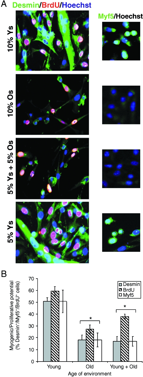
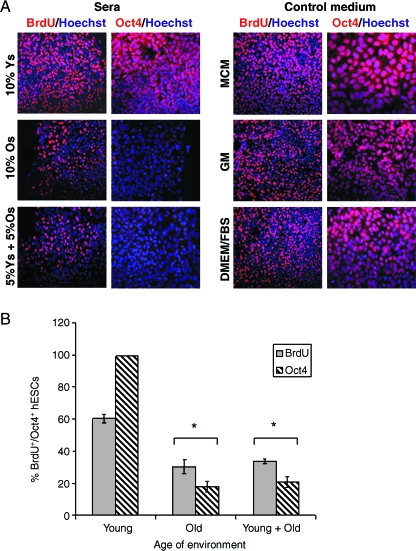
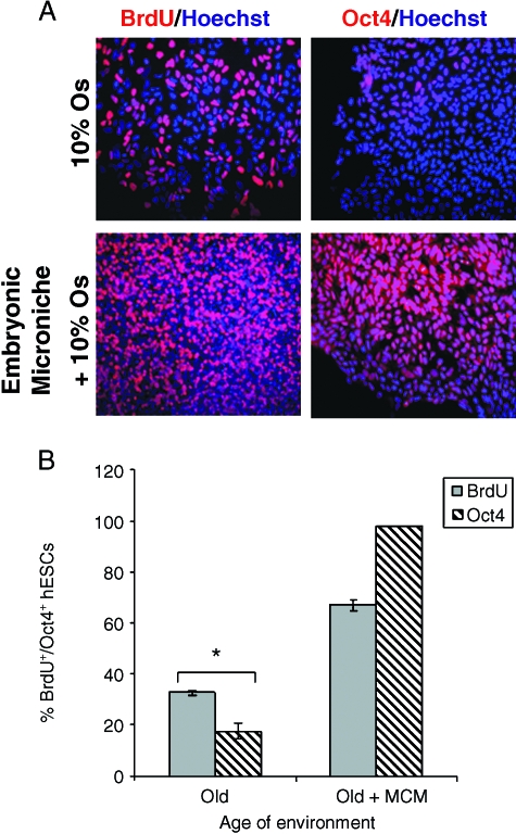
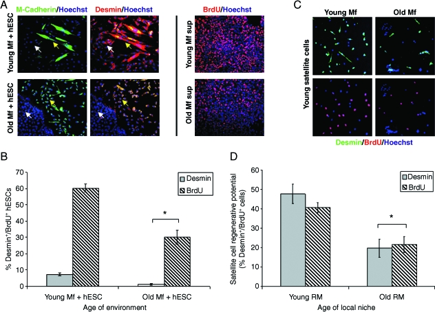
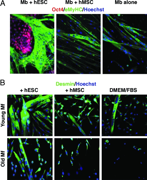
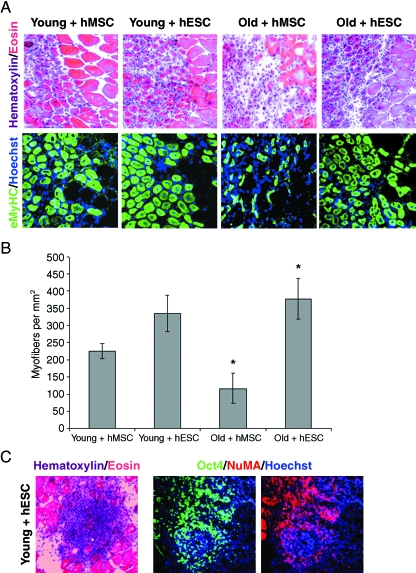
This work uncovers novel mechanisms of aging within stem cell niches that are evolutionarily conserved between mice and humans and affect both embryonic and adult stem cells. Specifically, we have examined the effects of aged muscle and systemic niches on key molecular identifiers of regenerative potential of human embryonic stem cells (hESCs) and post-natal muscle stem cells (satellite cells). Our results reveal that aged differentiated niches dominantly inhibit the expression of Oct4 in hESCs and Myf-5 in activated satellite cells, and reduce proliferation and myogenic differentiation of both embryonic and tissue-specific adult stem cells (ASCs). Therefore, despite their general neoorganogenesis potential, the ability of hESCs, and the more differentiated myogenic ASCs to contribute to tissue repair in the old will be greatly restricted due to the conserved inhibitory influence of aged differentiated niches. Significantly, this work establishes that hESC-derived factors enhance the regenerative potential of both young and, importantly, aged muscle stem cells in vitro and in vivo; thus, suggesting that the regenerative outcome of stem cell-based replacement therapies will be determined by a balance between negative influences of aged tissues on transplanted cells and positive effects of embryonic cells on the endogenous regenerative capacity. Comprehensively, this work points toward novel venues for in situ restoration of tissue repair in the old and identifies critical determinants of successful cell-replacement therapies for aged degenerating organs.


Figures

Fig. 1
Fig. 1
The age of sera determined the regenerative potential of satellite cells. (A) Young satellite cells were cultured either in 5% or 10% young (Young), 10% old (Old), or in a 5%+ 5% mouse sera combination (young + old). Cells were analyzed by immunofluorescence microscopy, using anti-BrdU (red), antidesmin (green) or anti-Myf5 antibodies (green, small panels). Similar results are shown for Pax7 immunodetection (Supplementary Fig. S1). Hoechst (blue) labeled nuclei. (B) Three independent experiments were quantified [300 young myofibers per experiment] as percentage of desmin+/Myf5+/BrdU+ de novo generated cells for each age and culture condition. On average, two to three fewer cells were generated when cultured in the presence of old. Shown are identical microscope fields at ×40 magnification. At least three independent experiments produced similar results. (*) indicates P≤ 0.001 as compared to young sera.
Fig. 2
Fig. 2
The regenerative potential of embryonic stem cells was negatively affected by aged mouse sera. (A) hESCs were cultured in MCM with 10% young (young) or old (old) mouse serum, or in three control media: MCM without mouse sera; GM (myoblast medium of Ham's F10 with 20% FBS) and DMEM/FBS (hESC differentiation medium of DMEM with 10% FBS). BrdU was added for the last 2 h of culture to measure the rate of cell proliferation. Immunodetection assays were performed for BrdU (red), Oct4 (red), and Ki67 (Supplementary Fig. S2). Hoechst (blue) labels nuclei. A high rate of hESC proliferation and Oct4 expression is displayed in all control media and in the presence of young mouse serum. In contrast, hESC proliferation and Oct4 expression are inhibited in the presence of old mouse serum, either alone or when mixed with young serum. MCM with mouse sera at 5% gave results similar to those observed with 10% young mouse sera or in control media (Supplementary Fig. S3). (B) Three independent experiments yielded similar results and were quantified as percentage of BrdU+ and Oct4+ cells for each culture condition. * indicates P < 0.001 as compared to young serum.
Fig. 3
Fig. 3
Embryonic stem cells produce youthful microniche in culture. (A) As opposed to immediate exposure to old mouse serum after passaging (10% old), preculturing of hESCs for 24 h in feeder-free conditions, e.g., Matrigel™ + MCM, prior to replacing MCM with MCM + 10% old mouse sera, resulted in continuously high BrdU incorporation and Oct4 expression (embryonic microniche + 10% old). BrdU was added for the last 2 h of culture to measure the rate of cell proliferation. Immunodetection of BrdU and Oct4 (both in red) was performed as described in Experimental procedures. Hoechst (blue) labels nuclei. (B) Three independent experiments yielded similar results and were quantified as percentage of BrdU+/Oct4+ for each condition. * indicates P < 0.001 as compared to ‘old + MCM’.
Fig. 4
Fig. 4
Aged muscle niche inhibits the regenerative potential of hESCs and satellite cells. (A) Immunodetection of a mouse-specific M-cadherin (green) or desmin (red; both human and mouse proteins are detected) revealed that hESCs underwent muscle lineage differentiation when co-cultured with young, but not old myofibers. The myogenic progeny of hESCs appears M-cadherin/desmin+ (white arrow in young), as opposed to M-cadherin/desmin hESCs that lack myogenic commitment (white arrow in old). M-cadherin+/desmin+ cells are the myogenic progeny of mouse satellite cells (yellow arrows). To assess the effects of secreted factors produced by young vs. old myofibers on the rate of hESC proliferation, transient, 2 h BrdU incorporation was examined in hESCs cultured for 48 h with supernatants produced by heterochronic myofiber explants (See Experimental procedures for details). As compared to young myofiber-derived supernatants (young myofiber supernant), exposure to old myofiber-derived supernatants (old myofiber supernant) inhibited hESCs proliferation, as judged by BrdU immunodetection (red). As expected, the rate of hESCs proliferation was high in control media (shown in Fig. 2). Hoechst (blue) labels nuclei in all experiments. Quantification of desmin+/BrdU+ hESCs in direct myofiber cocultures, or with muscle supernatants, is shown in (B). * indicates P≤ 0.001 as compared to young. (C) Transwell co-cultures between purified young satellite cells and myofibers isolated from uninjured young (young myofiber) and old (old myofiber) muscle demonstrated that satellite cell regenerative myogenic capacity was inhibited by the aged differentiated muscle. Myogenic potential was determined by the ability of satellite cells to generate proliferating desmin+ myoblasts (immunodetection shown in green) and by rate of proliferation (2 h BrdU incorporation; immunodetection shown in red). (D) Satellite cell regenerative potential was quantified as percentage of desmin+/BrdU+ cells for transwell co-cultures with young or old uninjured myofibers (i.e., RM, resting muscle). n = 3; * indicates P≤ 0.05 as compared to young.
Fig. 5
Fig. 5
In vitro co-culture with hESCs enhanced myogenesis of mouse cells. (A) 1 × 10⁵ hESCs or control hMSCs were co-cultured with 5 × 10⁶ primary mouse myoblasts. hESCs expressing Oct4 (immunodetection shown in red) dramatically enhanced myotube formation of co-cultured mouse myoblasts (immunodetection of eMyHC is shown in green), as compared to co-cultures between mouse myoblasts and human mesenchymal stem cells (Mb + hMSCs) or myoblasts alone (Mb alone). Experiments were carried out in myoblast differentiation medium. Hoechst (blue) labels nuclei throughout this figure. (B) 1 × 10⁵ hESCs or control hMSCs were co-cultured with young or old myofiber-associated satellite cells, as described in Experimental procedures. Co-culture with hESCs (myofiber + hESC), but not hMSCs (myofiber + hMSC) or control medium (DMEM/FBS), greatly enhanced the myogenic potential of both young and old myofiber-associated satellite cells, based on immunodetection of percentage of desmin+ de novo generated myoblasts and multinucleated myotubes. These experiments were carried out in GM. Shown are myogenic responses of mouse cells only, judged by lack of immunoreactivity to human-specific/hESC-specific antigens, such as NuMA and Oct4; and presence of mouse-specific immunoreactivity, e.g., M-cadherin (not shown). Both young and old myofiber associated satellite cells exhibited considerable myogenic improvement over control conditions. n = 3.
Fig. 6
Fig. 6
Skeletal muscle regeneration following hESC transplantation is a balance between the inhibitory influence of aged niches and the rejuvenating effects of hESCs. Young and old tibialis anterior and gastrocnemius muscles were injured by cardiotoxin injection. hESCs or hMSCs were transplanted at the site of injury and were analyzed by cryosectioning at Day 5 after injury (as described in Experimental procedures). (A) Newly regenerated myofibers were detected using eMyHC-specific antibody (green) and staining with H&E. In H&E staining, newly regenerated areas contain smaller, immature myofibers with centrally located nuclei. Uninjured myofibers are much larger, by comparison, with peripherally restricted nuclei. Poorly regenerated areas lack new myofibers and contain areas of fibrosis and inflammation. eMyHC immunodetection is specific for regenerating areas of muscle only. Both assays showed dramatic enhancement of muscle regeneration in ‘old + hESC’ vs. ‘old + hMSC’. Regeneration improvement was also seen in young + hESC, as compared to young + hMSC. (B) Quantification of muscle regeneration was performed by analyzing the density of newly formed myofibers per mm2 of injury site, which is the volume that typically covers the whole injured area. Multiple, 10 µm H&E sections were examined through the entire volume of injury in multiple, independently injured muscles. n = 20; * indicates P < 0.001 (‘old + hMSC’ compared to young + hMSC and ‘old + hMSC’ compared to ‘old + hESC’. (C) H&E and immunofluoresence staining for Oct4, and a human-specific antibody to NuMA, revealed the failure of hESCs to expand or persist in old, but the presence of hESCs in young muscle at 5 days post-transplantation. Hoechst (blue) labels nuclei.

Similar articles

Cited by

References

    1. Blyszczuk P, Czyz J, Kania G, Wagner M, Roll U, St-Onge L, Wobus AM. Expression of Pax4 in embryonic stem cells promotes differentiation of nestin-positive progenitor and insulin-producing cells. Proc. Natl Acad. Sci. USA. 2003;100:998–1003. - PMC - PubMed
    1. Carlson BM, Faulkner JA. Muscle transplantation between young and old rats: age of host determines recovery. Am. J. Physiol. 1989;256:C1262–C1266. - PubMed
    1. Collins CA, Olsen I, Zammit PS, Heslop L, Petrie A, Partridge TA, Morgan JE. Stem cell function, self-renewal, and behavioral heterogeneity of cells from the adult muscle satellite cell niche. Cell. 2005;122:289–301. - PubMed
    1. Conboy IM, Rando TA. The regulation of Notch signaling controls satellite cell activation and cell fate determination in postnatal myogenesis. Dev. Cell. 2002;3:397–409. - PubMed
    1. Conboy IM, Conboy MJ, Smythe GM, Rando TA. Notch-mediated restoration of regenerative potential to aged muscle. Science. 2003;302:1575–1577. - PubMed

Publication types

Substances