. 2007 Apr;14(4):283-92.
doi: 10.1111/j.1365-2893.2006.00796.x.

T-cell epitope mapping of ORF2 and ORF3 proteins of human hepatitis E virus

Affiliations

T-cell epitope mapping of ORF2 and ORF3 proteins of human hepatitis E virus

R Aggarwal et al. J Viral Hepat. 2007 Apr.

Abstract

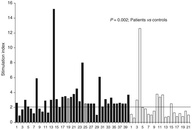
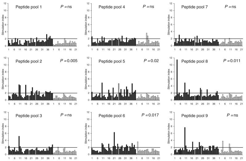
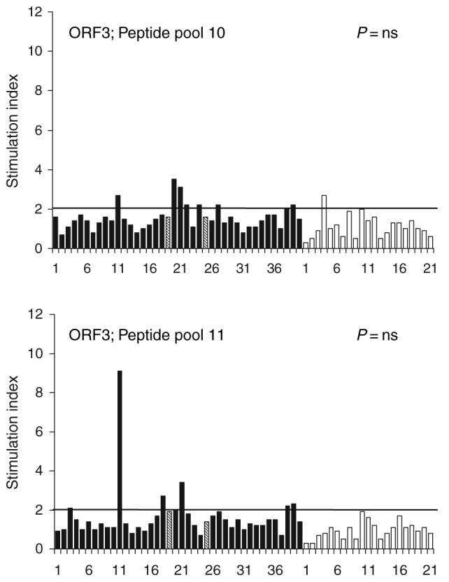
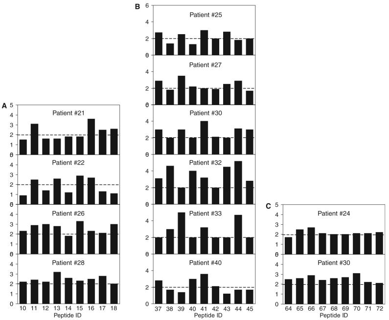
Little data are available on cellular immune responses during infection with hepatitis E virus (HEV). We therefore mapped CD4 T-cell epitopes in open reading frame (ORF)2 and ORF3 proteins of HEV using lymphocyte proliferation assays and overlapping peptide libraries. Proliferation of peripheral blood mononuclear cells from 40 patients with acute hepatitis E and 21 healthy controls with recombinant HEV ORF2 protein or pools of overlapping HEV ORF2/ORF3 peptides was measured. HLA-DQB1 and HLA-DRB1 alleles were also determined. Mononuclear cells from patients with hepatitis E more often showed significant proliferation on stimulation with recombinant ORF2 protein than controls (32/40 vs 7/21), and had higher median (range) stimulation indices [2.6 (0.9-15.2) vs 1.3 (0.6-12.9)]. Peptide pools corresponding to amino acids 73-156, 289-372, 361-444 and 505-588 of HEV ORF2 protein were associated with significant proliferation. Individual peptides in these pools did not show a clear pattern of stimulation. HEV ORF3 peptide pools did not induce proliferative responses. Lymphocyte proliferation in response to the peptide pool corresponding to amino acids 289-372 of HEV ORF2 protein was associated with presence of HLA-DRB1 allele 010X. These data on mapping of T-cell epitopes in HEV proteins may prove useful for designing HEV vaccines and for studying the immunopathogenesis of hepatitis E.


Figures

Fig. 1
Fig. 1
Results of lymphocyte proliferation assays in response to recombinant HEV ORF2 protein among patients with acute hepatitis E (solid bars) and healthy controls (empty bars). The bars indicate stimulation indices in the presence of recombinant hepatitis E virus ORF2 protein (10 μg/mL). The horizontal line indicates the cut-off (2.0), above which stimulation indices were considered as significant. Stimulation indices in patients were higher than those among controls (medians 2.6 and 1.3, respectively; P = 0.002, Wilcoxon’s rank sum test). The numbers on X-axis represent serial numbers for patients and controls. Hatched lines represent the two patients (19 and 25) who had fulminant hepatic failure.
Fig. 2
Fig. 2
Results of lymphocyte proliferation assays in response to pools of peptides corresponding to HEV ORF2 protein among patients with acute hepatitis E (solid bars) and healthy controls (empty bars). The bars indicate stimulation indices in the presence of pools of nine 20-mer peptides (10 μg/mL each). The horizontal line indicates the cut-off of 2.0, above which stimulation indices were considered as significant. For four pools (pools 2, 5, 6 and 8), the proportion of patients with significant stimulation was higher among patients than in controls. The numbers on X-axes represent serial numbers for patient and controls. Hatched lines represent the two patients (19 and 25) who had fulminant hepatic failure.
Fig. 3
Fig. 3
Results of lymphocyte proliferation assays in response to pools of peptides corresponding to HEV ORF3 protein among patients with acute hepatitis E (solid bars) and healthy controls (empty bars). The bars indicate stimulation indices in the presence of pools of seven 20-mer peptides (10 μg/mL each) each. The horizontal line indicates the cut-off of 2.0, above which stimulation indices were considered as significant. For both pools, the proportion of patients with significant stimulation was similar among patients and controls. The numbers on X-axes represent serial numbers for patient and controls. Hatched lines represent the two patients (19 and 25) who had fulminant hepatic failure.
Fig. 4
Fig. 4
Results of lymphocyte proliferation assays in response to individual peptides contained in pools 2, 5 and 8 of peptides corresponding to HEV ORF2 protein among patients with acute hepatitis E. The bars indicate stimulation indices in the presence of individual 20-mer peptides (10 μg/mL). Panel A indicates stimulation indices with peptides 10 to 18 in four patients, panel B those with peptides 37 to 45 in six patients with acute hepatitis E and panel C those with peptides 64 to 72 in two patients with acute hepatitis E. The horizontal line indicates the cut-off of 2.0, above which stimulation indices were considered as significant.

References

    1. Aggarwal R, Krawczynski K. Hepatitis E: an overview and recent advances in clinical and laboratory research. J Gastroenterol Hepatol. 2000;15:9–20. - PubMed
    1. Krawczynski K, Aggarwal R. Hepatitis E. In: Schiff ER, Sorrell MF, Maddrey WC, editors. Schiff’s Diseases of the Liver. 9th edn. Philadelphia, PA: Lippincott-Raven; 2002. pp. 877–889.
    1. Naik SR, Aggarwal R, Salunke PN, Mehrotra NN. A large waterborne viral hepatitis E epidemic in Kanpur, India. Bull World Health Organ. 1992;70:597–604. - PMC - PubMed
    1. Aggarwal R, Naik SR. Hepatitis E: intrafamilial transmission versus waterborne spread. J Hepatol. 1994;22:718–723. - PubMed
    1. Somani SK, Aggarwal R, Naik SR, Srivastava S, Naik S. A serological study of intra-familial spread from patients with sporadic hepatitis E virus infection. J Viral Hepat. 2003;10:446–449. - PubMed

Publication types

MeSH terms