

Review
doi: 10.1186/ar2135.
Bone morphogenetic proteins in destructive and remodeling arthritis
Affiliations
- PMID: 17381828
- PMCID: PMC1906799
- DOI: 10.1186/ar2135
Item in Clipboard
Review
Bone morphogenetic proteins in destructive and remodeling arthritis
Arthritis Res Ther.
2007.
Abstract
Joint destruction and tissue responses determine the outcome of chronic arthritis. Joint inflammation and damage are often the dominant clinical presentation. However, in some arthritic diseases, in particular the spondyloarthritides, joint remodeling is a prominent feature, with new cartilage and bone formation leading to ankylosis and contributing to loss of function. A role for bone morphogenetic proteins in joint remodeling has been demonstrated in the formation of both enthesophytes and osteophytes. Data from genetic models support a role for bone morphogenetic protein signaling in cartilage homeostasis. Finally, this signaling pathway is likely to play a steering role in the synovium.
Figures
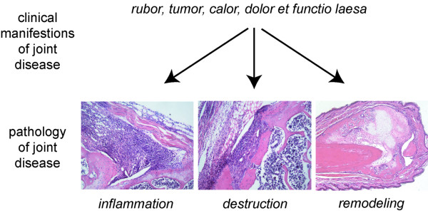
The signs and symptoms of arthritis are caused by distinct processes in the joint. Synovitis with extensive inflammation is characteristic. Formation of pannus tissue and activation of osteoclasts contributes to joint destruction. Tissue remodeling is characterized by new cartilage and bone formation eventually leading to ankylosis. The images presented were obtained from mice with methylated bovine serum albumin-induced arthritis (inflammation and destruction) and from mice with spontaneous ankylosing enthesitis (remodeling).
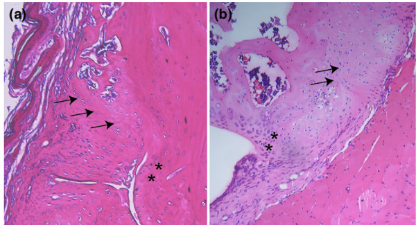
Enthesophytes and osteophytes are different. (a) The enthesophyte originates from the insertion of capsule and tendons (arrows). The chondrosynovial border of the articular cartilage is not involved (asterisks). (b) Osteophyte originating from the border of the articular cartilage (asterisks). In contrast, the enthesis is normal (arrows).
References
Publication types
MeSH terms
Substances
LinkOut - more resources
Full Text Sources
Medical
