

Ubp43 gene expression is required for normal Isg15 expression and fetal development
- PMID: 17381847
- PMCID: PMC1852108
- DOI: 10.1186/1477-7827-5-13
Ubp43 gene expression is required for normal Isg15 expression and fetal development
Abstract
Background: Isg15 covalently modifies murine endometrial proteins in response to early pregnancy. Isg15 can also be severed from targeted proteins by a specific protease called Ubp43 (Usp18). Mice lacking Ubp43 (null) form increased conjugated Isg15 in response to interferon. The Isg15 system has not been examined in chorioallantoic placenta (CP) or mesometrial (MM) components of implantation sites beyond 9.5 days post coitum (dpc). It was hypothesized that deletion of Ubp43 would cause disregulation of Isg15 in implantation sites, and that this would affect pregnancy rates.
Methods: Heterozygous (het) Ubp43 mice were mated and MM and CP implantation sites were collected on 12.5 and 17.5 days post-coitum (dpc).
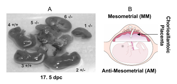
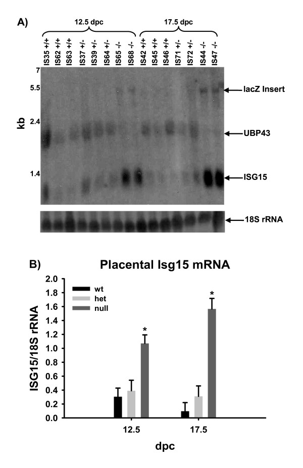
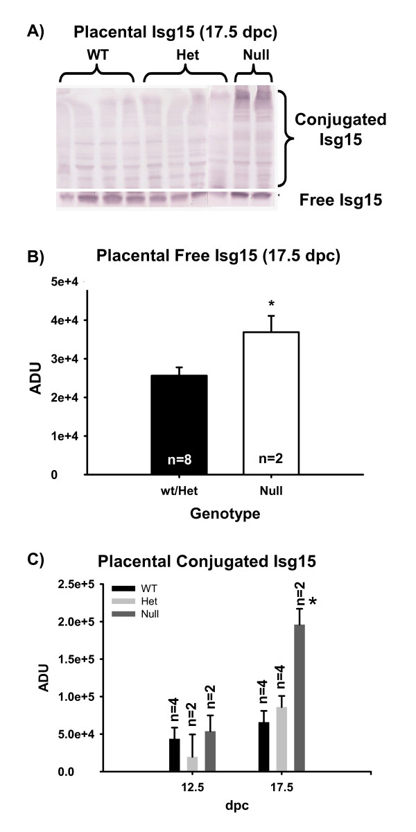
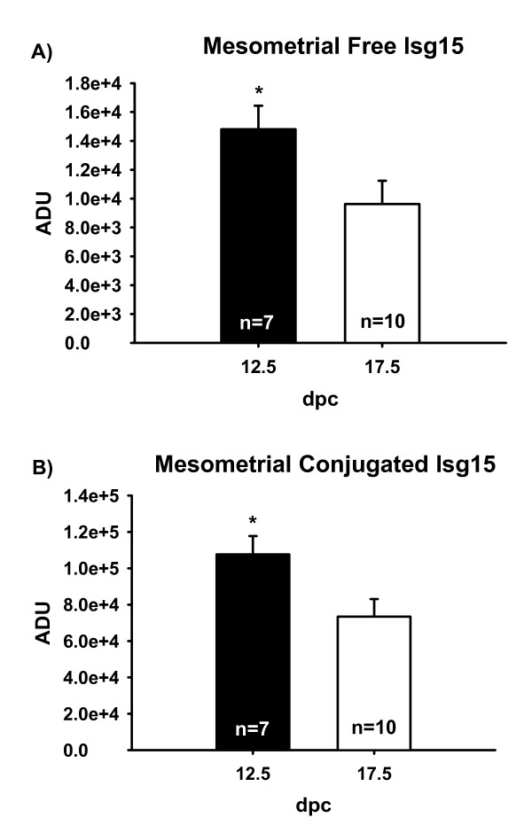
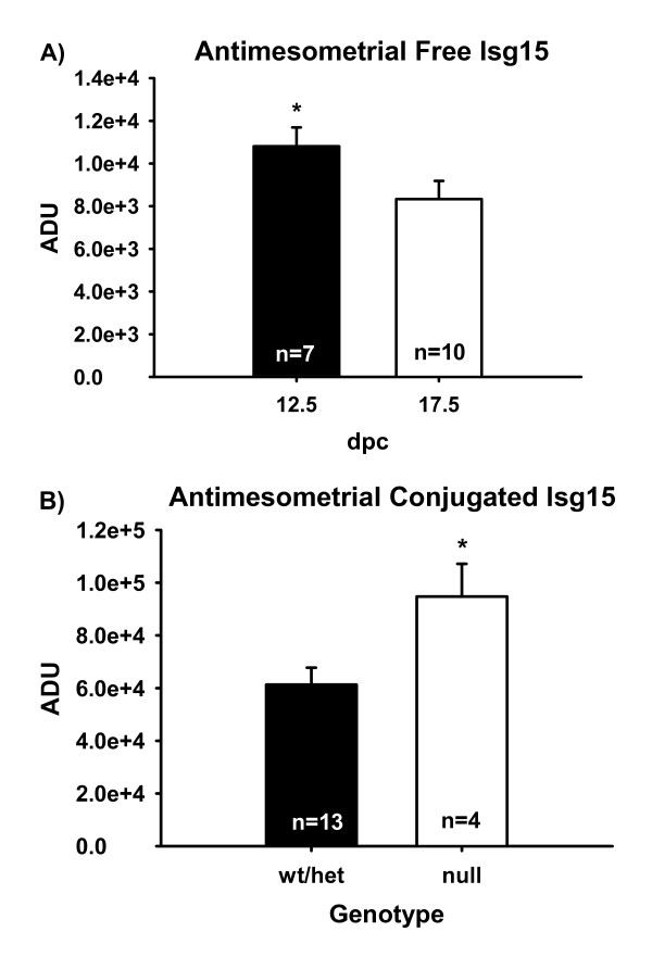
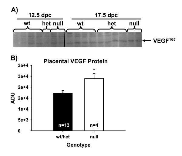
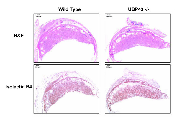
Results: Free and conjugated Isg15 were greater on 12.5 versus 17.5 dpc in MM. Free and conjugated Isg15 were also present in CP, but did not differ due to genotype on 12.5 dpc. However, null CP had greater free and conjugated Isg15 when compared to het/wt on 17.5 dpc. Null progeny died in utero with fetal genotype ratios (wt:het:null) of 2:5:1 on 12.5 and 2:2:1 on 17.5 dpc. Implantation sites were disrupted within the junctional zone and spongiotrophoblast, contained less vasculature based on lectin B4 staining and contained greater Isg15 mRNA and VEGF protein in Ubp43 null when compared to wt placenta.
Conclusion: It is concluded that Isg15 and its conjugates are present in implantation sites during mid to late gestation and that deletion of Ubp43 causes an increase in free and conjugated Isg15 at the feto-maternal interface. Also, under mixed genetic background, deletion of Ubp43 results in fetal death.
Figures
Similar articles
-
Deletion of the Isg15 gene results in up-regulation of decidual cell survival genes and down-regulation of adhesion genes: implication for regulation by IL-1beta.Endocrinology. 2010 Sep;151(9):4527-36. doi: 10.1210/en.2010-0166. Epub 2010 Jul 21. Endocrinology. 2010. PMID: 20660068
-
Embryo mortality in Isg15-/- mice is exacerbated by environmental stress.Biol Reprod. 2015 Feb;92(2):36. doi: 10.1095/biolreprod.114.122002. Epub 2014 Dec 10. Biol Reprod. 2015. PMID: 25505199
-
Reexamination of the role of ubiquitin-like modifier ISG15 in the phenotype of UBP43-deficient mice.Mol Cell Biol. 2005 Dec;25(24):11030-4. doi: 10.1128/MCB.25.24.11030-11034.2005. Mol Cell Biol. 2005. PMID: 16314524 Free PMC article.
-
Interferon-stimulated gene 15 and the protein ISGylation system.J Interferon Cytokine Res. 2011 Jan;31(1):119-30. doi: 10.1089/jir.2010.0110. Epub 2010 Dec 29. J Interferon Cytokine Res. 2011. PMID: 21190487 Free PMC article. Review.
-
The ISG15/USP18 ubiquitin-like pathway (ISGylation system) in hepatitis C virus infection and resistance to interferon therapy.Int J Biochem Cell Biol. 2011 Oct;43(10):1427-31. doi: 10.1016/j.biocel.2011.06.006. Epub 2011 Jun 16. Int J Biochem Cell Biol. 2011. PMID: 21704181 Review.
Cited by
-
Gene expression profiling in the submandibular gland, stomach, and duodenum of CAVI-deficient mice.Transgenic Res. 2011 Jun;20(3):675-98. doi: 10.1007/s11248-010-9441-2. Epub 2010 Sep 11. Transgenic Res. 2011. PMID: 20835760
-
Do molecular signals from the conceptus influence endometrium decidualization in rodents?J Exp Zool B Mol Dev Evol. 2009 Dec 15;312(8):797-816. doi: 10.1002/jez.b.21308. J Exp Zool B Mol Dev Evol. 2009. PMID: 19551814 Free PMC article. Review.
-
Evidence for the ubiquitin protease UBP43 as an antineoplastic target.Mol Cancer Ther. 2012 Sep;11(9):1968-77. doi: 10.1158/1535-7163.MCT-12-0248. Epub 2012 Jul 2. Mol Cancer Ther. 2012. PMID: 22752428 Free PMC article.
-
Epstein-Barr virus independent dysregulation of UBP43 expression alters interferon-stimulated gene expression in Burkitt lymphoma.PLoS One. 2009 Jun 24;4(6):e6023. doi: 10.1371/journal.pone.0006023. PLoS One. 2009. PMID: 19551150 Free PMC article.
-
Protein ISGylation: a posttranslational modification with implications for malignant neoplasms.Explor Target Antitumor Ther. 2023;4(4):699-715. doi: 10.37349/etat.2023.00162. Epub 2023 Aug 31. Explor Target Antitumor Ther. 2023. PMID: 37711589 Free PMC article. Review.
References
-
- Austin KJ, Pru JK, Hansen TR. Complementary deoxyribonucleic acid sequence encoding bovine ubiquitin-cross reactive protein: a comparison with ubiquitn and a 15-kDa ubiquitin homolog. Endocrine. 1996;5:191–197. - PubMed
-
- Johnson GA, Austin KJ, Collins AM, Murdoch WJ, Hansen TR. Endometrial ISG17 mRNA and a related mRNA are induced by interferon-tau and localized to glandular epithelial and stromal cells from pregnant cows. Endocrine. 1999;10:243–252. - PubMed
-
- Joyce MM, Hansen TR, Johnson GA. Interferon-stimulated gene 17 is expressed in the porcine uterus and may be critical to placental development across species. Biol Reprod. 2002;66:185. - PubMed
Publication types
MeSH terms
Substances
Grants and funding
LinkOut - more resources
Full Text Sources
Medical
Molecular Biology Databases
Miscellaneous
