. 2007 Apr 15;42(8):1290-302.
doi: 10.1016/j.freeradbiomed.2007.01.033. Epub 2007 Jan 24.

Aldose reductase mediates endotoxin-induced production of nitric oxide and cytotoxicity in murine macrophages

Affiliations

Aldose reductase mediates endotoxin-induced production of nitric oxide and cytotoxicity in murine macrophages

Kota V Ramana et al. Free Radic Biol Med. .

Abstract

Aldose reductase (AR) is a ubiquitously expressed protein with pleiotrophic roles as an efficient catalyst for the reduction of toxic lipid aldehydes and mediator of hyperglycemia, cytokine, and growth factor-induced redox-sensitive signals that cause secondary diabetic complications. Although AR inhibition has been shown to be protective against oxidative stress signals, the role of AR in regulating nitric oxide (NO) synthesis and NO-mediated apoptosis has not been elucidated to date. We therefore investigated the role of AR in regulating lipopolysaccharide (LPS)-induced NO synthesis and apoptosis in RAW 264.7 macrophages. Inhibition or RNA interference ablation of AR suppressed LPS-stimulated production of NO and overexpression of iNOS mRNA. Inhibition or ablation of AR also prevented the LPS-induced apoptosis, cell cycle arrest, activation of caspase-3, p38-MAPK, JNK, NF-kappaB, and AP1. In addition, AR inhibition prevented the LPS-induced down-regulation of Bcl-xl and up-regulation of Bax and Bak in macrophages. L-Arginine increased and L-NAME decreased the severity of cell death caused by LPS and AR inhibitors prevented it. Furthermore, inhibition of AR prevents cell death caused by HNE and GS-HNE, but not GS-DHN. Our findings for the first time suggest that AR-catalyzed lipid aldehyde-glutathione conjugates regulate the LPS-induced production of inflammatory marker NO and cytotoxicity in RAW 264.7 cells. Inhibition or ablation of AR activity may be a potential therapeutic target in endotoximia and other inflammatory diseases.


Figures
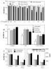
Fig. 1
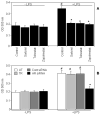
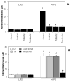
Fig. 1. Effect of AR inhibition/ablation on LPS-induced cell viability in macrophages
A) Growth –arrested macrophages were incubated without or with indicated AR inhibitors (10 μM) for 24 h and challenged with LPS (0–20 μg/ml) for another 24 h. B and C) cells were transfected with control or AR siRNA oligonucleotides followed by incubation with 10 μg/ml of LPS or 10 μg/ml LPS + 100 U/ml of IFN-γ for 24 h. A to C) The cell viability was determined by MTT assay and C) by cell counts as described in the Methods. The inset in Fig 1C shows the Western blot for AR protein expression. Data are expressed as Mean ± SEM (N = 4). *P < 0.001 as compared to LPS-treated cells #P < 0.001 control cells. UT, untransfected; TR, Transfection Reagent; Con; Scrambled SiRNA; AR siR; AR siRNA.
Fig. 2
Fig. 2. Effect of AR inhibition on LPS-induced apoptosis in macrophages
Growth –arrested macrophages were A) incubated without or with indicated AR inhibitors (10 μM), B) transfected with control or AR siRNA oligonucleotides and both A and B were challenged with LPS (10 μg/ml) for 24 h. The apoptosis was determined by measuring nucleosomal degradation using ELISA kit. All the data are expressed as Mean ± SEM (N = 4). *P < 0.001 as compared to LPS-treated cells #P < 0.001 control cells.
Fig. 3
Fig. 3. Effect of AR inhibition on LPS-induced morphological changes in macrophages
Growth –arrested macrophages were incubated without or with indicated AR inhibitors (10 μM), and challenged with LPS (10 μg/ml) for 24 h. Subsequently, the cells were stained with A) 5 μg/ml of Hoechst 33342 and B) 1 μg/ml of propidium iodide for 30 min at 4°C to morphologically identify apoptotic cells (blue) and Necrotic cells (red), respectively. C) The cells shown in bright filed. All the pictures were taken at 40X magnifications using a Nikon epifluorescence microscope.
Fig. 4
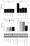
Fig. 4. Effect of AR inhibition/ablation on LPS-induced activation of caspase-3 in macrophages
Growth –arrested macrophages were A & C) incubated without or with indicated AR inhibitors (10 μM), B) transfected with control or AR siRNA oligonucleotides and challenged with LPS (10 μg/ml) for 24 h. The caspase-3 activity was determined by A and B) in vitro ELISA kit and C) in situ PARP cleavage by Western blot using antibodies against PARP as described in the Methods. All the data are expressed as Mean ± SEM (N = 4). *P < 0.001 as compared to LPS-treated cells #P < 0.001 control cells.
Fig. 5
Fig. 5. AR inhibition prevents LPS-induced activation of p38 MAPK and JNK in macrophages
Growth –arrested macrophages were incubated without or with AR inhibitor, sorbinil (10 μM), and challenged with LPS (1 μg/ml) for indicated times. A-D) Western blots were developed using antibodies against A) phospho-p38 MAPK, B) total p38 MAPK, C) phospho-JNK and D) total JNK. Antigen-antibody complex was detected by enhanced chemiluminescence.
Fig. 6
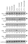
Fig. 6. AR inhibition prevents LPS-induced regulation of BCl-2 family of proteins
Growth –arrested macrophages were incubated without or with AR inhibitors (10 μM), and challenged with LPS (10 μg/ml) for 24 h. A–F) Western blots were developed using antibodies against A) Bcl-2, B) Bcl-xl, C) Bax, D) Bak, E) Bad and F) GAPDH. Antigen-antibody complex was detected by enhanced chemiluminescence.
Fig. 7
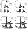
Fig. 7. Inhibition of AR prevents LPS-induced arrest of synthesis phase of cell cycle in macrophages
Growth-arrested macrophages were pre-incubated with sorbinil or tolrestat or carrier for 24 h followed by stimulation with of LPS for 24 h and cell cycle analysis was performed by FACS.
Fig. 8
Fig. 8. AR inhibition prevents LPS-induced nitric oxide production in macrophages
Growth-arrested macrophages were A) pre-incubated with AR inhibitors or carrier for 24 h, B) transfected with control or AR siRNA oligonucleotides and both A and B were challenged with LPS (1 μg/ml) for 24 h. In the culture media Nitrate/nitrite levels were measured by using specific ELISA kits as described in the methods. All the data are expressed as Mean ± SEM (N = 4). *P < 0.001 as compared to LPS-treated cells #P < 0.001 control cells.
Fig. 9
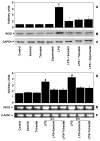
Fig. 9. AR inhibition prevents LPS-induced iNOS expression in macrophages
Growth-arrested macrophages were pre-incubated with AR inhibitors or carrier for 24 h followed by the incubation with LPS (1 μg/ml) for additional 24 h. In the cell extracts iNOS expression was measured by B) Western blot analysis and E) RT-PCR as described in the methods. A & D) Densitometric analysis of B and E. C & F) Loading controls. All the data are expressed as Mean ± SEM (N = 4). *P < 0.001 as compared to LPS-treated cells #P < 0.001 control cells.
Fig. 10
Fig. 10. AR inhibition prevents LPS-induced activation of NF-κB and AP1 in macrophages
Growth-arrested macrophages were A & C) pre-incubated with AR inhibitors or carrier for 24 h and B & D) transfected with control or AR siRNA oligonucleotides. Subsequently all the four (A–D) were incubated with LPS (1 μg/ml) for 2 h. Equal amounts of nuclear extracts were subjected to EMSA for A & B) NF-κB and C & D) AP1 as described in the Methods.
Fig. 11
Fig. 11. Effect of NO in LPS-induced macrophage viability
Growth-arrested macrophages were pre-incubated with sorbinil for 24 h followed by incubation with L-arginine or L-NAME for 2 h. Subsequently the macrophages were incubated with LPS (10 □g/ml) for 24h. The cell viability was determined by MTT assay as described in the methods. All the data are expressed as Mean ± SEM (N = 4). *P < 0.001 as compared to LPS-treated cells, #P < 0.001 control cells and ##P < 0.01 as compared to LPS-treated cells.
Fig. 12
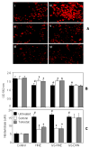
Fig. 12. Effect of AR inhibition on GS-aldehydes –induced cytotoxicity in macrophages
A) The growth arrested macrophages with or without sorbinil or tolrestat were treated with Dihydroethidium (hydroehidine) for 15 min followed by LPS for 60 min. The fluorescence intensity was evaluated under Nikon Epifluorescence microscope (40X magnification). I) Control, II) Sorbinil, III) Tolrestat, IV) LPS, V) LPS+Sorbinil and VI) LPS+Tolrestat. B and C) Growth-arrested macrophages were pre-incubated with AR inhibitors or carrier for 24 h followed by the incubation with HNE, GS-HNE-ester or GS-DHN-ester (1 μM) for additional 24 h. B) The cell viability was determined by MTT assay and C) the nitrate/nitrite levels were measured by using specific ELISA kits as described in the methods. All the data are expressed as Mean ± SEM (N = 4). *P < 0.001 as compared to aldehyde-treated cells #P < 0.01 control cells.

Similar articles

Cited by

References

    1. Raetz CR, Ulevitch RJ, Wright SD, Sibley CH, Ding A, Nathan CF. Gram-negative endotoxin: an extraordinary lipid with profound effects on eukaryotic signal transduction. FASEB J. 1991;5:2652–2660. - PubMed
    1. Heine H, Rietschel ET, Ulmer AJ. The biology of endotoxin. Mol Biotechnol. 2001;19:279–296. - PubMed
    1. Lopez-Bojorquez LN, Dehesa AZ, Reyes-Teran G. Molecular mechanisms involved in the pathogenesis of septic shock. Arch Med Res. 2004;35:465–479. - PubMed
    1. Wolkow PP. Involvement and dual effects of nitric oxide in septic shock. Inflamm Res. 1998;47:52–166. - PubMed
    1. Baumgarten G, Knuefermann P, Schuhmacher G, Vervolgyi V, von Rappard J, Dreiner U, Fink K, Djoufack C, Hoeft A, Grohe C, Knowlton AA, Meyer R. Toll-like receptor 4, nitric oxide, and myocardial depression in endotoxemia. Shock. 2006;25:43–49. - PubMed

Publication types