Review
. 2007 Mar;1771(3):353-404.
doi: 10.1016/j.bbalip.2007.01.015. Epub 2007 Feb 6.

Synthesis and function of membrane phosphoinositides in budding yeast, Saccharomyces cerevisiae

Affiliations
Review

Synthesis and function of membrane phosphoinositides in budding yeast, Saccharomyces cerevisiae

Thomas Strahl et al. Biochim Biophys Acta. 2007 Mar.

Abstract

It is now well appreciated that derivatives of phosphatidylinositol (PtdIns) are key regulators of many cellular processes in eukaryotes. Of particular interest are phosphoinositides (mono- and polyphosphorylated adducts to the inositol ring in PtdIns), which are located at the cytoplasmic face of cellular membranes. Phosphoinositides serve both a structural and a signaling role via their recruitment of proteins that contain phosphoinositide-binding domains. Phosphoinositides also have a role as precursors of several types of second messengers for certain intracellular signaling pathways. Realization of the importance of phosphoinositides has brought increased attention to characterization of the enzymes that regulate their synthesis, interconversion, and turnover. Here we review the current state of our knowledge about the properties and regulation of the ATP-dependent lipid kinases responsible for synthesis of phosphoinositides and also the additional temporal and spatial controls exerted by the phosphatases and a phospholipase that act on phosphoinositides in yeast.


Figures

Figure 1
Figure 1. Chemical structure of phosphatidylinositol (PtdIns)
The inositol headgroup is esterified to inorganic phosphate, which is in turn esterified to diacylglycerol thereby forming a phosphodiester link. In vivo the lipid portion of the molecule is directly inserted into the lipid bilayer, whereas the hydrophilic headgroup protrudes into the cytosol on the cytoplasmic surface of biological membranes. This leaves the hydroxyls in positions D3, D4 and D5 accessible for cytoplasmic and membrane-bound enzymes such as lipid kinases and phosphatases.
Figure 2
Figure 2. Interconversion of phosphoinositides in yeast
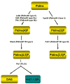
(A) Phosphoinositide kinase reactions in S. cerevisiae. The individual steps in the synthesis of phosphoinositides catalyzed by lipid kinases, as well as the breakdown of PtdIns[4,5]P2 by phosphoinositide-specific phospholipase C (Plc1) are depicted. All phosphoinositide kinases that have been identified in yeast exhibit specificity for a single substrate. The phosphorylation of PtdIns to PtdIns[4]P can be catalyzed by three different enzymes, namely Lsb6, Pik1 and Stt4, whereas all other reactions are catalyzed only by one kinase. The classification for each enzyme is also illustrated. (B) Phosphoinositide phosphatase reactions in yeast. In contrast to the phosphoinositide kinases, the lipid phosphatases of S. cerevisiae show a high degree of redundancy, as most of them dephosphorylate a number of substrates with relatively little specificity. Only Ymr1, Fig 4, Inp51/Sjl1 and Inp54 are highly selective for a single substrate, whereas Inp52/Sjl2 and Inp53/Sjl3 can convert most phosphoinositides that have been identified in yeast to PtdIns [235].
Figure 3
Figure 3. Domain structure of the PtdIns kinases and their accessory proteins in yeast
The different conserved domains are represented by colored boxes and their positions within the primary sequences of the proteins are also indicated. Domains that share a significant degree of homology, such as the lipid kinase unique (LKU) domain and the catalytic domains of Vps34, Pik1 and Stt4, are shown in the same color. (A) Domain structure of the PtdIns 3-kinase Vps34 and its accessory protein Vps15. The interaction of both proteins is mediated by a short 28 residues-spanning region in the COOH-terminal catalytic domain of Vps34 and the three HEAT repeat motifs located in the center of Vps15. The C2 domain of Vps34 and the WD-40 motif of Vps15 are also depicted. (B) Domain structure of the PtdIns 4-kinases of yeast and their accessory factors, Sfk1 and Frq1. The bipartite catalytic domain of Lsb6 is shown in a different color, as it is unrelated to those of the other lipid kinases in yeast. The region of Stt4 that is sandwiched between its LKU and catalytic domains is similar to the pleckstrin homology (PH) domains found in other proteins. The non-functional EF-hand motif in Frq1, which contains substitutions in key residues necessary for Ca2+ coordination, is indicated with an asterisk and demarcated with a different color than the other three EF-hand motis. C2, C2 domain; EF, EF-hand motif; HEAT, HEAT repeat motif; LKU, lipid kinase unique domain; NHD, novel homology domain; PH, pleckstrin homology-like domain; TM, transmembrane domain; WD-40, WD-40 motif.
Figure 4
Figure 4. Vacuolar protein sorting of carboxypeptidase Y (CPY)
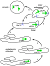
CPY is synthesized at the ER as an inactive precursor, prepro-CPY. Upon release into the ER lumen its NH2-terminal signal sequence is removed proteolytically, thereby generating pro-CPY (1). Subsequently, it undergoes NH2-linked glycosylation to become the ER-modified 67 kDa form p1CPY (2). In a next step p1CPY is transported to the Golgi apparatus where it receives further oligosaccharide modifications yielding another intermediate form, the 69 kDa p2CPY (3). In the late Golgi p2CPY is bound by Vps10, which serves as a transport receptor for regulated trafficking to a prevacuolar endosomal compartment (PVC), away from secretory cargo destined for the plasma membrane (4). In the PVC p2CPY is released from Vps10, which itself is recycled back to the Golgi apparatus for another round of receptor-mediated transport between these two compartments (5). Following its delivery to the vacuole p2CPY is cleaved by lumenal proteases into the active, mature 61 kDa form mCPY (6).
Figure 5
Figure 5. Vps34 containing PtdIns 3-kinase complexes
The yeast PtdIns 3-kinase, Vps34, forms two different multi-subunit complexes (Complex I and Complex II), which function in distinct biological processes, namely the autophagy/cytosol-to-vacuole transport (Cvt) and vacuolar protein sorting (Vps) pathways. Both complexes share the Vps15, Vps34 and Vps30 subunits, whereas Atg14 is limited to complex I and Vps38 is specific for complex II. Vps15 is NH2-myristoylated, and, hence, mediates the recruitment of complexes I and II to the pre-autophagosomal structure (PAS) and the prevacuolar endosomal compartment (PVC), respectively. In addition, Atg14 is essential for the recruitment of complex I to the PAS, whereas complex II localizes to the PVC in a manner that does not depend on the presence of Vps38. Phosphorylation of Vps34 by Vps15, which plays a role in the interaction of both proteins, as well as in the binding of Vps34 to Atg14 and Vps38, is also depicted.
Figure 6
Figure 6. Schematic representation of the autophagy and cytosol-to-vacuole (Cvt) pathways in yeast
Aminopeptidase 1 (Ape1), which is synthesized as an inactive precursor (prApe1) in the cytoplasm follows both pathways for its delivery to the vacuolar lumen where it is proteolytically processed to yield the mature form of the enzyme (mApe1), is shown as a model substrate. The commitment of yeast cells to either pathway depends on nutrient conditions. In both pathways, autophagy and cytosol-to-vacuole transport, cytosol and in the case of autophagy also entire organelles are engulfed and sequestered by a double membrane. The initial step of this process, which also referred to as vesicle nucleation, takes place at the pre-autophagosomal structure (PAS). Factors known to be involved in the function of this organelle, including the PtdIns 3-kinase Vps34, are also shown. Upon completion, autophagosomes and Cvt vesicles are targeted to and fuse with the vacuolar membrane. Ultimately, the resulting autophagic and Cvt bodies are degraded in the vacuolar lumen.
Figure 7
Figure 7. Schematic representation of the cellular functions of Pik1
Pik1 and Frq1 both localize to Golgi membranes, where they, together with Sec14, play a role in the regulation of secretion and, in sporulating cells, prospore formation. Pik1 also shuttles between the cytosol and the nucleus in a manner that depends on the karyopherins Kap95 and Msn5. One possible nuclear function of Pik1 is the regulation of one or more processes required for successful completion of meiosis.
Figure 8
Figure 8. Schematic representation of the cellular functions of Stt4
Stt4 localizes to the plasma membrane in a manner that requires its accessory protein Sfk1. Stt4 functions upstream of both Pkc1 and Cla4 in the regulation of polarized growth and the cell wall integrity/heat shock response pathways. Furthermore, evidence for a role of Stt4 in aminophospholipid transport from the ER has been obtained [209].
Figure 9
Figure 9. Domain structure of the PtdInsP-kinases and their accessory proteins in yeast
Conserved regions are shown as colored boxes and homologous regions are depicted in the same color. CCT, chaperonin containing T-complex homology region; FYVE, Fab1, YGL023, Vps27, and EEA1 domain; HEAT, HEAT repeat motif; TM, transmembrane domain.
Figure 10
Figure 10. Schematic representation of the cellular functions of Mss4
(A) Regulation of the subcellular localization and activity of Mss4 and its role in the cell wall integrity pathway. Nucleocytoplasmic shuttling of Mss4 depends on Kap123 and Bcp1, whereas its recruitment to the plasma membrane involves phosphorylation by the casein kinase I isoforms Yck1 and Yck2. At the plasma membrane Mss4 acts downstream of the PtdIns 4-kinase Stt4 and upstream of the Rho GEF Rom2, which directly binds to PtdIns[4,5]P2 generated by Mss4. In concert with one of the cell surface sensors that function in cell wall integrity sensing such as Wsc1, Rom2 stimulates nucleotide exchange on Rho1, which in turn activates the β-1,3-glucan synthase Fks1. Another effector of activated Rho1-GTP is the serine/threonine kinase Pkc1, which serves as an upstream activator of the Slt2/Mpk1 MAP kinase cascade. Ultimately this signaling cascade leads to changes in the transcriptional output of a number of genes including FKS1. (B) Model for the regulation of the actin cytoskeleton by Mss4. Slm1 and Slm2 directly bind PtdIns[4,5]P2 generated by Mss4 at the plasma membrane, which is itself stimulated by the input of Cmd1. Furthermore, both proteins are subject to phosphorylation by Tor2, a component of the multisubunit TORC2 complex. Both inputs, phosphorylation by TORC2 and PtdIns[4,5]P2 binding are required for proper localization of Slm1/2 to the plasma membrane and the regulation of the actin cytoskeleton. Other downstream effectors of Mss4 that regulate the polarization of the actin cytoskeleton include the protein kinase Pkc1 and the formin Bni1.
Figure 11
Figure 11. Schematic representation of the cellular functions of Fab1
Fab1 acts in concert with its activators Vac7 and Vac14 at vacuolar/endosomal membranes, where it converts PtdIns[3]P generated by Vps34 to PtdIns[3,5]P2. This process is temporarily upregulated during conditions of hyperosmotic stress, in a manner that also requires the PtdIns[3,5]P2-specific phosphatase Fig 4. The downstream effectors of PtdIns[3,5]P2 include Ent3 and Ent5, as well as Atg18 and function in the regulation of vacuole acidification, the MVB pathway and vacuole to MVB membrane retrieval.
Figure 12
Figure 12. Domain structure of the phosphoinositide phosphatases in yeast
Conserved regions and other functional motifs are shown as colored boxes. GRAM, glucosyltransferase, Rab-like GTPase activator and myotubularin domain. The catalytically non-functional Sac1-like domain in Inp51/Sjl1, which contains substitutions in key active site residues [235], is demarcated with a different color and marked with an asterisk to distinguish it from the catalytically-competent Sac1-like domains present in Sac1, Fig 4, and the other two synaptojanin-like lipid phosphatases, Inp52/Sjl2 and Inp53/Sjl3. Although the Sac1-like domain of Inp51/Sjl1 may not hydrolyze any given phosphoinositide, it may contribute to physiological function of this phosphoinositide 5-phospatase, e.g. by binding to phosphoinositides and thereby assisting in membrane targeting and localization of the enzyme.
Figure 13
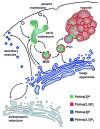
Figure 13. Model for the compartmentalization of phosphoinositides in yeast
The evidence accumulated to date indicates that different phosphoinositide species are generated in a compartment-specific manner and, hence, can be regarded as distinct markers for each organelle. For example, PtdIns[4]P is only detectable at membranes of the Golgi apparatus under normal conditions, and its aberrant accumulation in other locations, such as in the ER when cells carry mutations in genes that affect its turnover, display a number of severe phenotypes directly affecting growth. The pool of PtdIns[4]P generated at the plasma membrane is most likely rapidly and fully converted into PtdIns[4,5]P2, which is the major phosphoinositide at this location. PtdIns[3]P is found predominantly at membranes of the endosomal system and in multivesicular bodies, where a fraction is converted to PtdIns[3,5]P2, which is also found on vacuolar membranes. The presence and physiological roles of the different phosphoinositides in the nucleus of S. cerevisiae still remains unknown.

Similar articles

Cited by

References

    1. Carpenter CL, Cantley LC. Phosphoinositide kinases. Current Opinion in Cell Biology. 1996;8:153–8. - PubMed
    1. Fruman DA, Meyers RE, Cantley LC. Phosphoinositide kinases. Annual Review of Biochemistry. 1998;67:481–507. - PubMed
    1. Majerus PW. Inositol phosphatases and kinases in cell signaling. Faseb Journal. 1997;11:A1297–A1297.
    1. Hughes WE, Cooke FT, Parker PJ. Sac phosphatase domain proteins. Biochemical Journal. 2000;350:337–352. - PMC - PubMed
    1. Majerus PW, Kisseleva MV, Norris FA. The role of phosphatases in inositol signaling reactions. Journal of Biological Chemistry. 1999;274:10669–10672. - PubMed

Publication types

MeSH terms

Substances

LinkOut - more resources