

Structural and functional recovery of electropermeabilized skeletal muscle in-vivo after treatment with surfactant poloxamer 188
- PMID: 17382288
- PMCID: PMC1919408
- DOI: 10.1016/j.bbamem.2007.01.012
Structural and functional recovery of electropermeabilized skeletal muscle in-vivo after treatment with surfactant poloxamer 188
Abstract
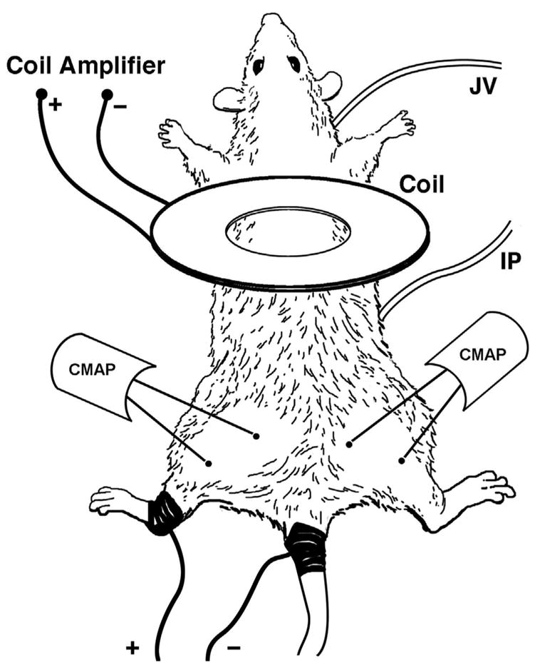
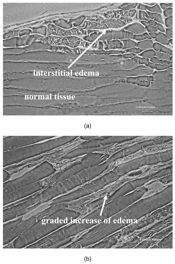
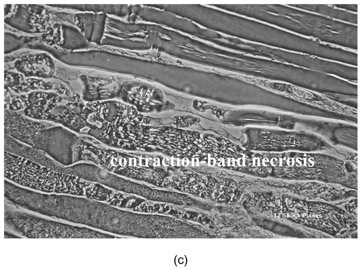
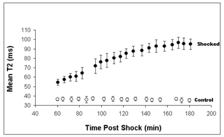
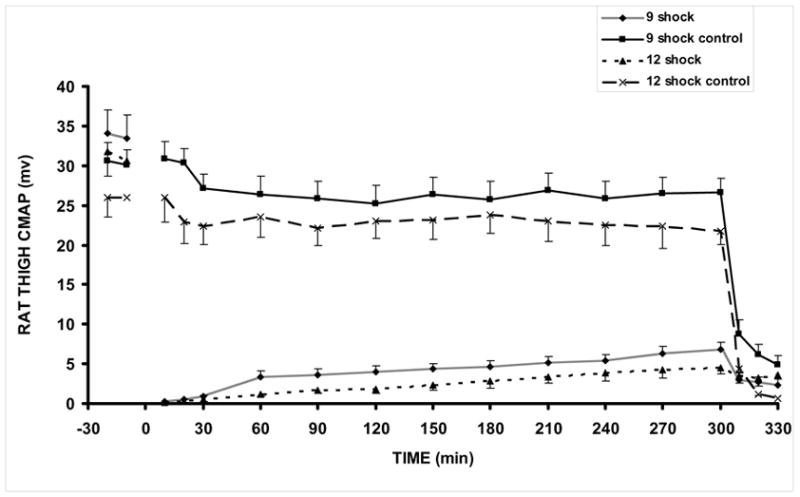
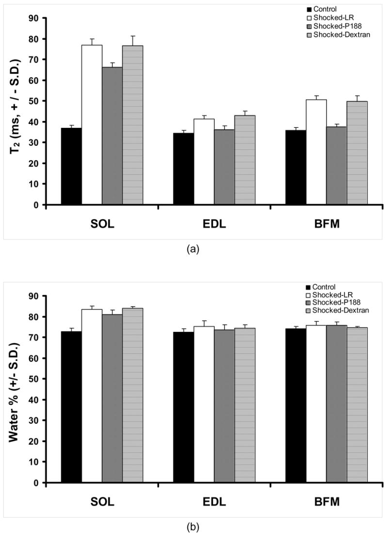
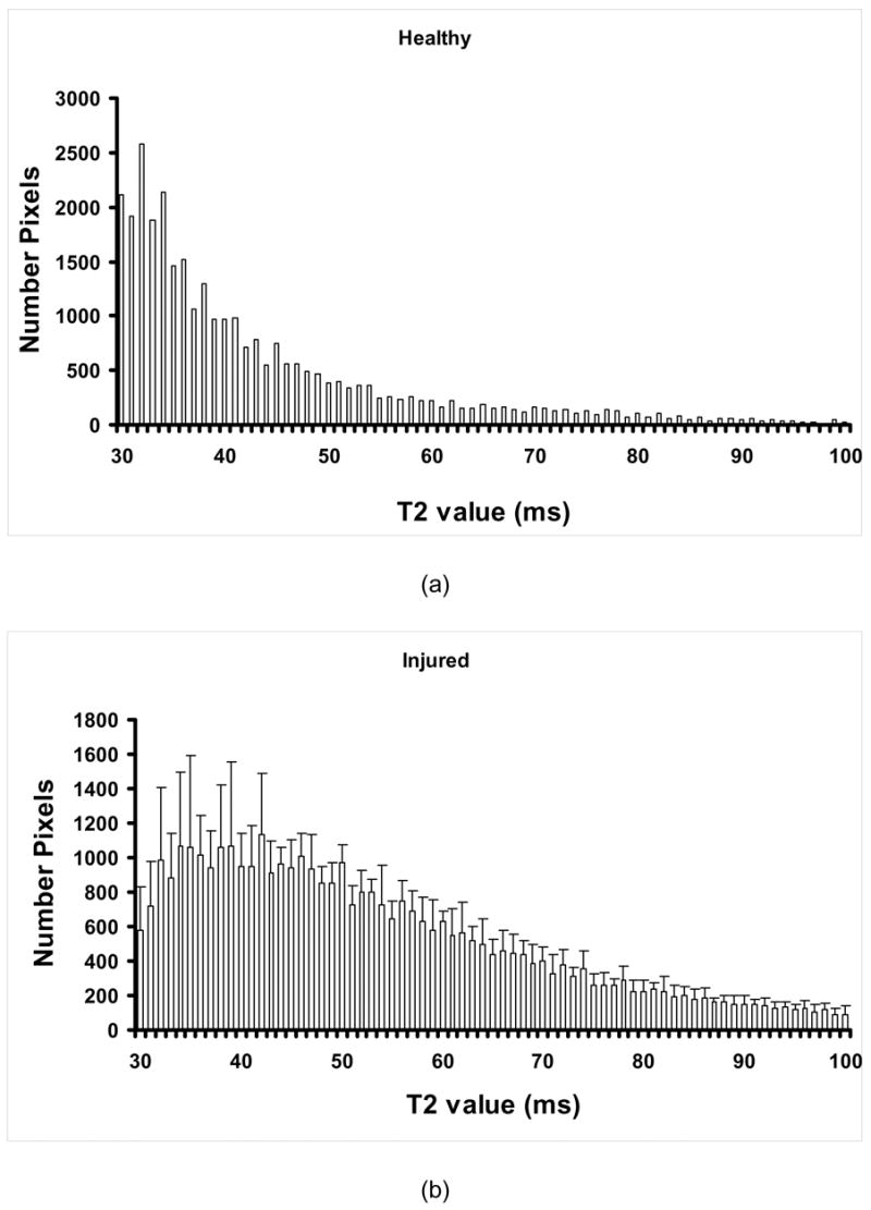
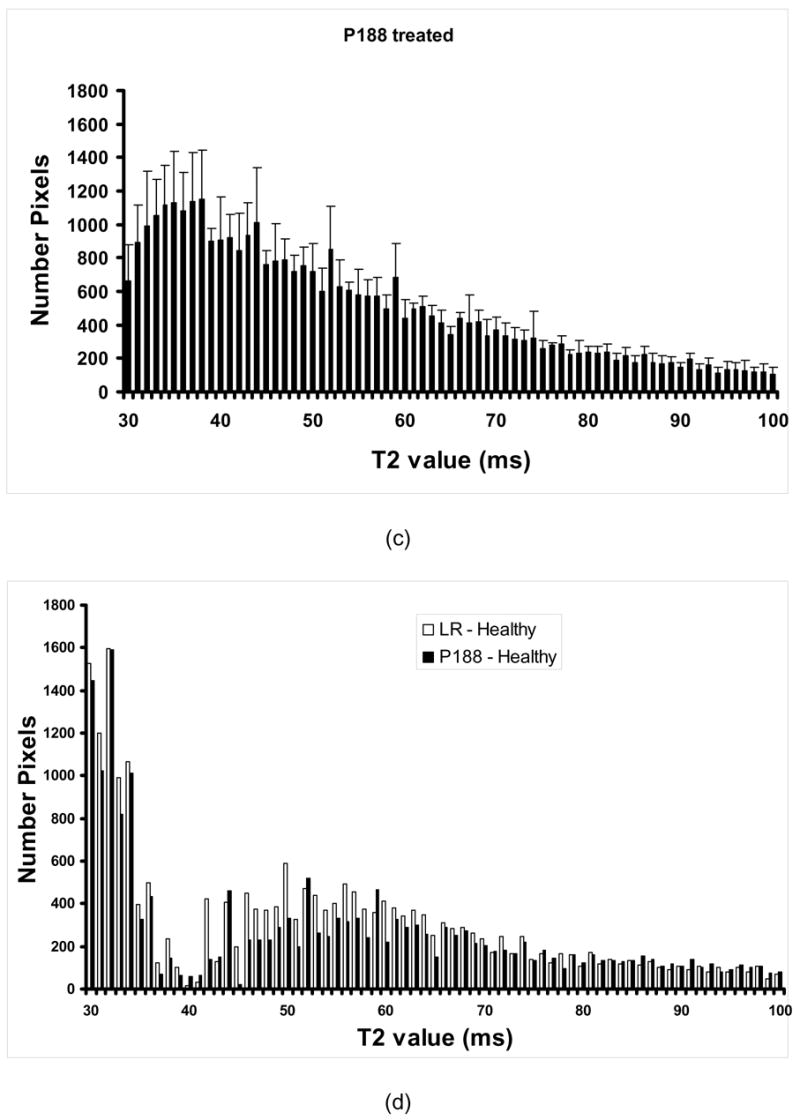
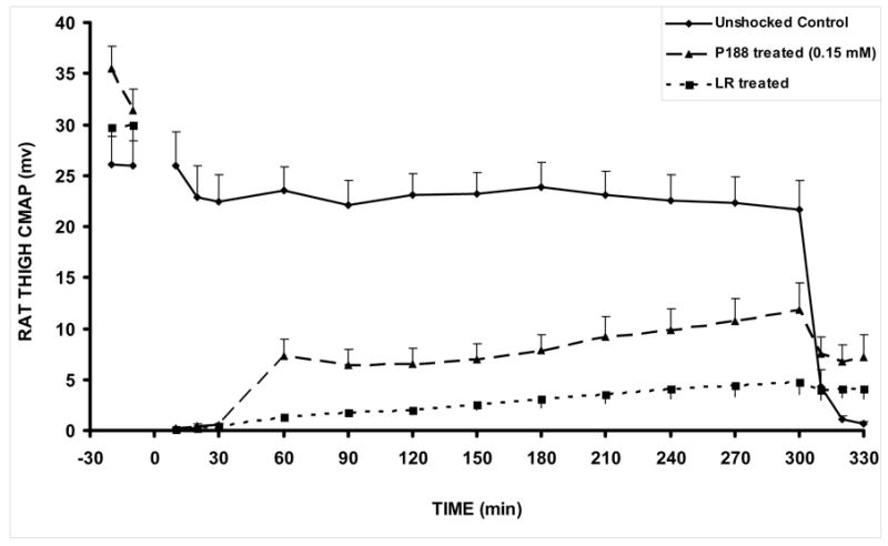
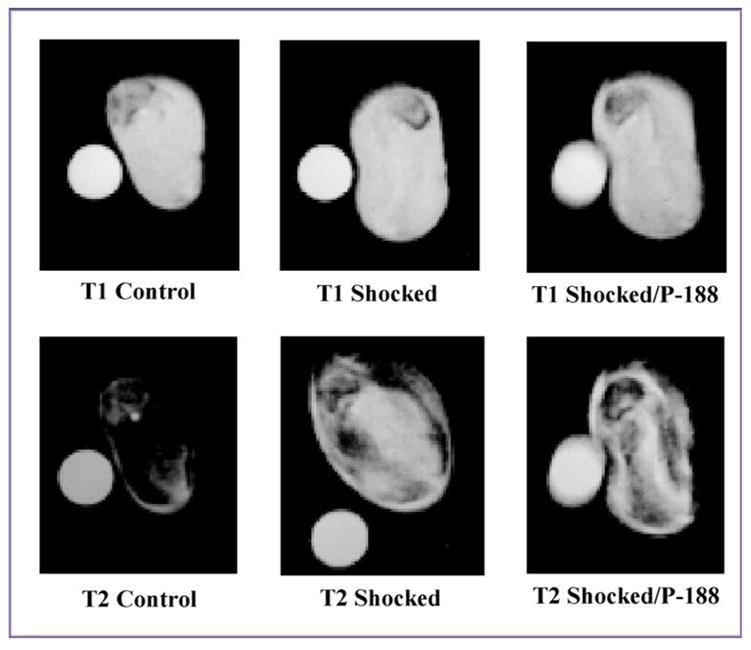
A critical requirement for cell survival after trauma is sealing of breaks in the cell membrane [M. Bier, S.M. Hammer, D.J. Canaday, R.C Lee, Kinetics of sealing for transient electropores in isolated mammalian skeletal muscle cells, Bioelectromagnetics 20 (1999) 194-201; R.C. Lee, D.C. Gaylor, D. Bhatt, D.A. Israel, Role of cell membrane rupture in the pathogenesis of electrical trauma, J. Surg. Res. 44 (1988) 709-719; R.C. Lee, J.F. Burke, E.G. Cravalho (Eds.), Electrical Trauma: The Pathophysiology, Manifestations, and Clinical Management, Cambridge University Press, 1992; B.I. Tropea, R.C. Lee, Thermal injury kinetics in electrical trauma, J. Biomech. Engr. 114 (1992) 241-250; F. Despa, D.P. Orgill, J. Newalder, R.C Lee, The relative thermal stability of tissue macromolecules and cellular structure in burn injury, Burns 31 (2005) 568-577; T.A. Block, J.N. Aarsvold, K.L. Matthews II, R.A. Mintzer, L.P. River, M. Capelli-Schellpfeffer, R.L. Wollman, S. Tripathi, C.T. Chen, R.C. Lee, The 1995 Lindberg Award. Nonthermally mediated muscle injury and necrosis in electrical trauma, J. Burn Care and Rehabil. 16 (1995) 581-588; K. Miyake, P.L. McNeil, Mechanical injury and repair of cells, Crit. Care Med. 31 (2003) S496-S501; R.C. Lee, L.P. River, F.S. Pan, R.L. Wollmann, Surfactant-induced sealing of electropermeabilized skeletal muscle membranes in vivo, Proc. Natl. Acad. Sci. 89 (1992) 4524-4528; J.D. Marks, C.Y. Pan, T. Bushell, W. Cromie, R.C. Lee, Amphiphilic, tri-block copolymers provide potent membrane-targeted neuroprotection, FASEB J. 15 (2001) 1107-1109; B. Greenebaum, K. Blossfield, J. Hannig, C.S. Carrillo, M.A. Beckett, R.R. Weichselbaum, R.C. Lee, Poloxamer 188 prevents acute necrosis of adult skeletal muscle cells following high-dose irradiation, Burns 30 (2004) 539-547; G. Serbest, J. Horwitz, K. Barbee, The effect of poloxamer-188 on neuronal cell recovery from mechanical injury, J. Neurotrauma 22 (2005) 119-132]. The triblock copolymer surfactant Poloxamer 188 (P188) is known to increase the cell survival after membrane electroporation [R.C. Lee, L.P. River, F.S. Pan, R.L. Wollmann, Surfactant-induced sealing of electropermeabilized skeletal muscle membranes in vivo, Proc. Natl. Acad. Sci. 89 (1992) 4524-4528; Z. Ababneh, H. Beloeil, C.B. Berde, G. Gambarota, S.E. Maier, R.V. Mulkern, Biexponential parametrization of T2 and diffusion decay curves in a rat muscle edema model: Decay curve components and water compartments, Magn. Reson. Med. 54 (2005) 524-531]. Here, we use a rat hind-limb model of electroporation injury to determine if the intravenous administration of P188 improves the recovery of the muscle function. Rat hind-limbs received a sequence of either 0, 3, 6, 9, or 12 electrical current pulses (2 A, 4 ms duration, 10 s duty cycle). Magnetic resonance imaging (MRI) analysis, muscle water content and compound muscle action potential (CMAP) amplitudes were compared. Electroporation injury manifested edema formation and depression of the CMAP amplitudes. P188 (one bolus of 1 mg/ml of blood) was administrated 30 or 60 min after injury. Animals receiving P188 exhibited reduced tissue edema (p<0.05) and increased CMAP amplitudes (p<0.03). By comparison, treatment with 10 kDa neutral dextran, which produces similar serum osmotic effects as P188, had no effect on post-electroporation recovery. Noteworthy, the present results suggest that a single intravenous dose of P188 is effective to restore the structural integrity of damaged tissues with intact circulation.
Figures
Similar articles
-
Poloxamer 188 prevents acute necrosis of adult skeletal muscle cells following high-dose irradiation.Burns. 2004 Sep;30(6):539-47. doi: 10.1016/j.burns.2004.02.009. Burns. 2004. PMID: 15302418
-
Surfactant-induced sealing of electropermeabilized skeletal muscle membranes in vivo.Proc Natl Acad Sci U S A. 1992 May 15;89(10):4524-8. doi: 10.1073/pnas.89.10.4524. Proc Natl Acad Sci U S A. 1992. PMID: 1584787 Free PMC article.
-
Mg ATP and antioxidants augment the radioprotective effect of surfactant copolymers.Health Phys. 2011 Dec;101(6):731-8. doi: 10.1097/HP.0b013e3182166759. Health Phys. 2011. PMID: 22048491 Free PMC article.
-
Functional tissue engineering for ligament healing: potential of antisense gene therapy.Ann Biomed Eng. 2004 Mar;32(3):342-51. doi: 10.1023/b:abme.0000017551.93144.1a. Ann Biomed Eng. 2004. PMID: 15098538 Review.
-
P188 Therapy in In Vitro Models of Traumatic Brain Injury.Int J Mol Sci. 2023 Feb 7;24(4):3334. doi: 10.3390/ijms24043334. Int J Mol Sci. 2023. PMID: 36834743 Free PMC article. Review.
Cited by
-
Spatial Distribution of PEO-PPO-PEO Block Copolymer and PEO Homopolymer in Lipid Bilayers.Langmuir. 2020 Apr 7;36(13):3393-3403. doi: 10.1021/acs.langmuir.9b03208. Epub 2020 Mar 27. Langmuir. 2020. PMID: 32216370 Free PMC article.
-
The Effect of Glucose and Poloxamer 188 on Red-Blood-Cell Aggregation.Metabolites. 2021 Dec 18;11(12):886. doi: 10.3390/metabo11120886. Metabolites. 2021. PMID: 34940644 Free PMC article.
-
Muscle membrane integrity in Duchenne muscular dystrophy: recent advances in copolymer-based muscle membrane stabilizers.Skelet Muscle. 2018 Oct 10;8(1):31. doi: 10.1186/s13395-018-0177-7. Skelet Muscle. 2018. PMID: 30305165 Free PMC article. Review.
-
Hyperamylinemia Increases IL-1β Synthesis in the Heart via Peroxidative Sarcolemmal Injury.Diabetes. 2016 Sep;65(9):2772-83. doi: 10.2337/db16-0044. Epub 2016 Jun 22. Diabetes. 2016. PMID: 27335231 Free PMC article.
-
Duchenne muscular dystrophy: disease mechanism and therapeutic strategies.Front Physiol. 2023 Jun 26;14:1183101. doi: 10.3389/fphys.2023.1183101. eCollection 2023. Front Physiol. 2023. PMID: 37435300 Free PMC article. Review.
References
-
- Bier M, Hammer SM, Canaday DJ, Lee RC. Kinetics of sealing for transient electropores in isolated mammalian skeletal muscle cells. Bioelectromagnetics. 1999;20:194–201. - PubMed
-
- Lee RC, Gaylor DC, Bhatt D, Israel DA. Role of cell membrane rupture in the pathogenesis of electrical trauma. J Surg Res. 1988;44:709–719. - PubMed
-
- Lee RC, Burke JF, Cravalho EG, editors. Electrical Trauma: The Pathophysiology, Manifestations, and Clinical Management. Cambridge University Press; 1992.
-
- Tropea BI, Lee RC. Thermal Injury Kinetics in Electrical Trauma. J Biomech Engr. 1992;114:241–250. - PubMed
-
- Despa F, Orgill DP, Newalder J, Lee RC. The Relative Thermal Stability of Tissue Macromolecules and Cellular Structure in Burn Injury. Burns. 2005;31:568–577. - PubMed
Publication types
MeSH terms
Substances
Grants and funding
LinkOut - more resources
Full Text Sources
Other Literature Sources
