Comparative Study
. 2007 May 1;305(1):333-46.
doi: 10.1016/j.ydbio.2007.02.014. Epub 2007 Feb 21.

Gcm2 is required for the differentiation and survival of parathyroid precursor cells in the parathyroid/thymus primordia

Affiliations
Comparative Study

Gcm2 is required for the differentiation and survival of parathyroid precursor cells in the parathyroid/thymus primordia

Zhijie Liu et al. Dev Biol. .

Abstract

The parathyroid glands develop with the thymus from bilateral common primordia that develop from the 3rd pharyngeal pouch endoderm in mouse embryos at about E11, each of which separates into one parathyroid gland and one thymus lobe by E13.5. Gcm2, a mouse ortholog of the Drosophila Glial Cells Missing gene, is expressed in the parathyroid-specific domains in the 3rd pouches from E9.5. The null mutation of Gcm2 causes aparathyroidism in the fetal and adult mouse and has been proposed to be a master regulator for parathyroid development. In order to study how Gcm2 functions in parathyroid development, we investigated the mechanism that causes the loss of parathyroids in Gcm2 null mutants. Analysis of the 3rd pouch-derived primordium in Gcm2-/- mutants showed the parathyroid-specific domain was present before E12.5 but underwent programmed cell death between E12 and 12.5. RNA and protein localization studies for parathyroid hormone (Pth) in wild-type embryos showed that the presumptive parathyroid domain in the parathyroid/thymus primordia started to transcribe Pth mRNA and produce PTH protein from E11.5 before the separation of parathyroid and thymus domains. However in Gcm2-/- mutants, the parathyroid-specific domain in the common primordium did not express Pth and could not maintain the expression of two other parathyroid marker genes, CasR and CCL21, although expression of these two genes was initiated. Marker gene analysis placed Gcm2 downstream of the known transcription and signaling pathways for parathyroid/thymus organogenesis. These results suggest that Gcm2 is not required for pouch patterning or to establish the parathyroid domain, but is required for differentiation and subsequent survival of parathyroid cells.


Figures
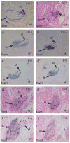
Fig. 1
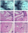
Fig. 1. Cell fate analysis of the 3rd pouch-derived parathyroid/thymus common primordia in wild-type and Gcm2−/− mutant embryos
Section in situ hybridization of Gcm2 on sections of wild-type embryos at E10.5 (A) and E11.5 (B) embryos is shown for comparison. Foxn1 in situ hybridization was performed on the sections from wild-type (C, E, G and I) and Gcm2−/− (D, F, H and J) embryos at E11.5 (C, D), E12 (E, F), E12.5 (G, H), and E13 (I, J) stages. Sections were cut in the sagittal plane. In all figures, anterior is up, and dorsal is to the right. Ages of embryos are indicated in the upper right corner of each panel. The thymus domain in (A, B), and the parathyroid domains in (C-J) are outlined. pt, parathyroid; th, thymus.
Fig. 2
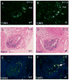
Fig. 2. TUNEL analysis of cell death in the parathyroid/thymus primordia in wild-type and Gcm2−/− mutant embryos
TUNEL was performed on a complete sagittal section series prepared from wild-type controls (A, E) and Gcm2−/− embryos (B, F) at E11.5 (A and B) and E12 (E and F) stages. Anterior is up, and dorsal is to the right. In (A-B), the common parathyroid/thymus primordium is outlined in white, and the 3rd pouch from which the primordium is undergoing separation by apoptosis is outlined in yellow (p3). At E12, in situ hybridization for Foxn1 to indicate the thymus domain (C, D) was performed on alternate sections with the sections used for TUNEL (E, F) to confirm that the location of the apoptotic cells are in the parathyroid domain in Gcm2 null mutant. The dorso-anterior parathyroid domain at E12 is indicated by a dashed line in C-F.
Fig. 3
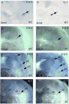
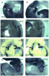
Fig. 3. The expression of CasR and CCL21 in wild-type and Gcm2−/− mutant embryos
Whole mount in situ hybridization for Gcm2 at E10.5 (A) and E11 (B) is shown for comparison. CCL21 (C, D) and CasR (E-H) expression is shown in wild-type (C, E, G) and Gcm2−/− (D, F, H) embryos at E10.5 (C-F). CasR expression is also shown at E11 (G, H). In all panels, dorsal is to the right, anterior is up. CasR expression was initiated at the dorsal sides of all four pouches (p1-p4) in the wild-type E10.5 embryo (E), and this expression was not affected by Gcm2 null mutation (F). The initiation of CCL21 expression at the dorsal side of 3rd pouch (C) partially required Gcm2 function (D). CasR expression was maintained only in the parathyroid domain at E11 in the wild-type (G), and this expression required Gcm2 function (H). a1, first arch; a2, second arch.
Fig. 4
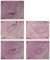
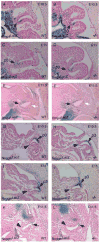
Fig. 4. Maintenance of CCL21 and CasR expression in the parathyroid domain at E11.5 requires Gcm2
Sections were cut at sagittal plane. In all figures, anterior is up, and dorsal is to the right. The common parathyroid/thymus primordium is outlined and the parathyroid domain is indicated with an arrow in (A, B, D). in situ hybridization for Gcm2 at E11.5 (A) is shown for comparison. CCL21 (B) and CasR (D) are restricted to the parathyroid domain at E11.5 in wild-type embryos. Neither is expressed in Gcm2 mutants (C, E). a1, first arch; a2, second arch.
Fig. 5
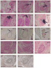
Fig. 5. The expression of Pth in wild-type and Gcm2−/− mutant embryos
Section in situ hybridization of Pth was performed on the sections prepared from the wild-type embryos at E10.5 (A), E11.5 (B), E12 (C), E12.5 (D), E13.5 (E), and E18.5 (F). Immunohistochemistry of PTH was performed on the sections prepared from the wild-type embryos at E11.5 (G), E12.5 (H), and E18.5 (I). The initiation of Pth mRNA and protein expression was not present in the Gcm2−/− mutants at E11.5 (J and M), and Pth mRNA and protein expression was also not present in late stages at E12 (K and N), and E12.5 (L). Sections were cut at sagittal plane. In all figures, anterior is up, and dorsal is to the right. Arrows in B-C indicate the Pth positive parathyroid domain. The common parathyroid/thymus primordium is outlined in (B, C, G, H, J-N). p3, third pouch; a1, first arch; a2, second arch; h, heart; th, thymus; tr, thyroid; pt, parathyroid.
Fig. 6
Fig. 6. Expression of Tbx1 in wild-type and Gcm2−/− mutant embryos
Whole mount (A, B) or paraffin section (C-F) in situ hybridization for Tbx1 at E10.5 (A-D) and E11.5 (E, F). In the wild-type, Tbx1 expression was present at the dorsal side of 3rd pouch at E10.5 (A, C) and at the parathyroid-specific domain in the parathyroid/thymus primordium at E11.5 (E). This expression is not affected by Gcm2 null mutation (B, D, F). Sections were cut in the saggital plane. Arrows in (E, F) indicate the parathyroid domain. p3, third pouch; a1, first arch; a2, second arch.
Fig. 7
Fig. 7. Expression of the Hoxa3-Pax1/9-Eya1 pathway in wild-type and Gcm2−/− mutant embryos
Whole mount in situ hybridization (A, B, C, D, G, and H) or LacZ staining (E and F) was performed on wild-type (A, C, E and G) and Gcm2−/− (B, D, F, and H) embryos at E10.5 for Hoxa3 (A and B), Pax1 (C and D), Pax9 (E and F), and Eya1 (G and H). The expression of these genes in the 3rd pouch was normal in Gcm2 mutants. The 3rd pouch is indicated in each panel (p3).
Fig. 8
Fig. 8. Expression of Shh and Ptc1 in the 3rd pouch of wild-type and Gcm2−/− mutant embryos
Section in situ hybridization for Shh (A and B) and Patched1 (C and D) was performed on transverse paraffin sections from wild-type (A and C) and Gcm2−/− (B and D) embryos at E10.5. The expression of these genes in the 3rd pouch was normal in Gcm2 mutants. Dorsal is up. p3, third pouch.
Fig. 9
Fig. 9. Expression of Bmp4 and Noggin in the 3rd pouch of wild-type and Gcm2−/− mutant embryos
Whole mount LacZ staining for Bmp4LacZ (A- F) and NogginLacZ (G-L) was performed on wild-type (A, C, E, G, I, and K) and Gcm2−/− (B, D, F, H, J, and L) at E10.5 (A, B, G and H), E11 (C, D, I and J), and E11.5 (E, F, K and L). In panels (A-F), arrows indicate Bmp4 expression restricted to the ventral/posterior thymus domain in the 3rd pouch (p3) and common primordium. In (E and F), the Bmp4-negative dorsal domain is indicated with a white arrow. In (G-L), arrows indicate dorsally restricted noggin domain. Arrowheads in (K and L) indicate the ventral thymus domain.

Similar articles

Cited by

References

    1. Ahn TG, Antonarakis SE, Kronenberg HM, Igarashi T, Levine MA. Familial isolated hypoparathyroidism: a molecular genetic analysis of 8 families with 23 affected persons. Medicine (Baltimore) 1986;65:73–81. - PubMed
    1. Balling R, Erben RG. From parathyroid to thymus, via glial cells. Nat Med. 2000;6:860–1. - PubMed
    1. Berg JP. A molecular switch for parathyroid cell differentiation. Eur J Endocrinol. 2002;146:281–2. - PubMed
    1. Blackburn CC, Augustine CL, Li R, Harvey RP, Malin MA, Boyd RL, Miller JF, Morahan G. The nu gene acts cell-autonomously and is required for differentiation of thymic epithelial progenitors. Proc Natl Acad Sci U S A. 1996;93:5742–6. - PMC - PubMed
    1. Blackburn CC, Manley NR. Developing a new paradigm for thymus organogenesis. Nat Rev Immunol. 2004;4:278–89. - PubMed

Publication types