

doi: 10.1016/j.advenzreg.2006.12.013.
Epub 2007 Mar 26.
Targeting the RAF/MEK/ERK, PI3K/AKT and p53 pathways in hematopoietic drug resistance
Affiliations
- PMID: 17382374
- PMCID: PMC2696319
- DOI: 10.1016/j.advenzreg.2006.12.013
Item in Clipboard
Targeting the RAF/MEK/ERK, PI3K/AKT and p53 pathways in hematopoietic drug resistance
Adv Enzyme Regul.
2007.
No abstract available
Figures
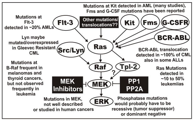
Sites of mutation which can result in activation of the Raf>MEK>ERK pathway. Mutations have been detected in Flt-3, Ras, Kit, Fms, G-CSFR, and at lower frequencies Raf-1 and B-Raf in AML. The BCR-ABL chromosomal translocation is present in virtually all CMLs and some ALLs. These mutations and chromosomal translocations could all result in activation of the Raf>MEK>ERK cascade. A ? is indicated in the connection between Tpl-2 and MEK. This is to indicate that there are other MEK activators besides Raf which can result in MEK activation and may confer sensitivity to MEK inhibitors. Mutations at phosphatase genes could also result in activation of this pathway although they would be predicted to be either tumor suppressor or dominant negative type mutations.
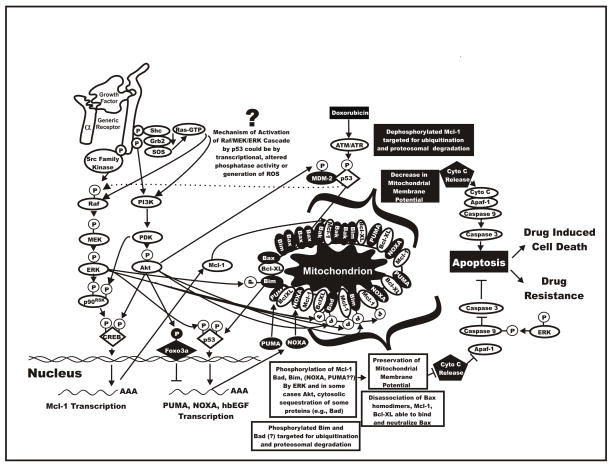
Overview of interactions between Raf>MEK>ERK, PI3K>Akt, p53 and apoptotic pathways resulting in drug resistance. The Raf>MEK>ERK and PI3K>Akt pathways can phosphorylate transcription factors which can stimulate gene transcription or apoptotic regulatory molecules which control the induction of apoptosis. Possibly through reactive oxygen species (ROS), doxorubicin can induce Raf>MEK>ERK. Doxorubicin can also activate p53 which can induce the transcription of molecules involved in the regulation of apoptosis. Heparin binding epidermal growth factor (hb-EGF) is a transcriptional target of p53 which could induce activation of the Raf>MEK>ERK cascade. Finally doxorubicin could induce p53 which alters the expression of phosphatases which could lead to prolonged ERK activation. Dysregulation of these cascades can result in the prevention of apoptosis and the induction of drug resistance.
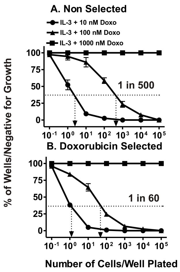
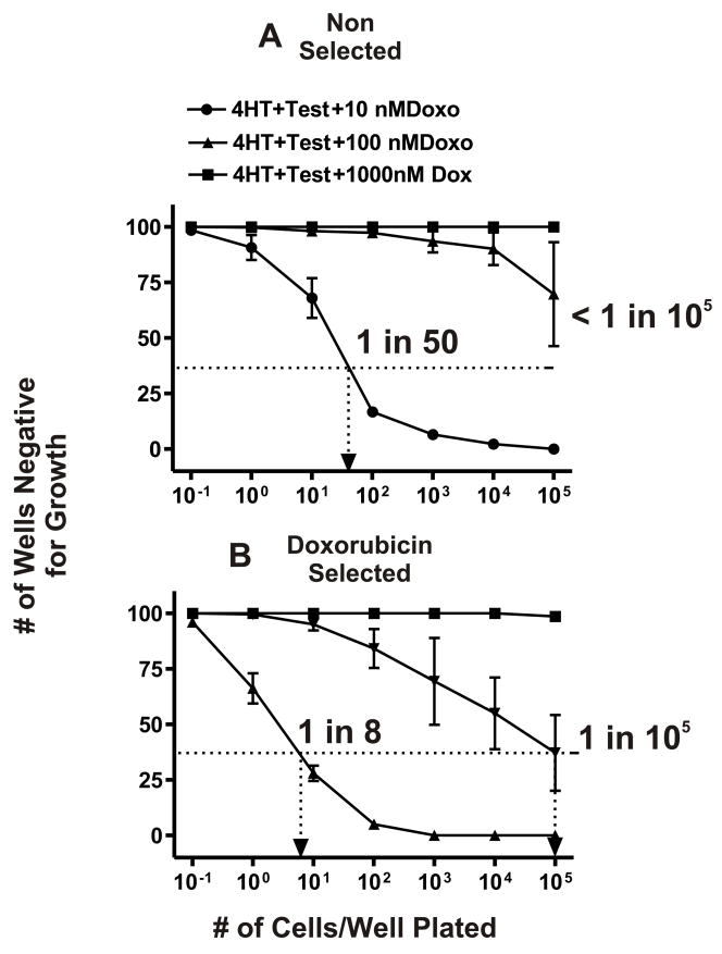
Isolation of doxorubicin resistant cells from FL5.12 and enhanced subcloning efficiency in doxorubicin. Limiting dilution analysis was performed in the presence of different concentrations of doxorubicin on the FL5.12 (Panel A) and doxorubicin resistant FL/Doxo (Panel B) cells. A dotted line is indicated at 37% of wells negative for growth from which the cloning efficiency can be estimated. These experiments have been performed 4 times and averaged together. Limiting dilution analysis with FL/Doxo-1 is presented in Panel B, similar results were observed with 2 other FL/Doxo clones.
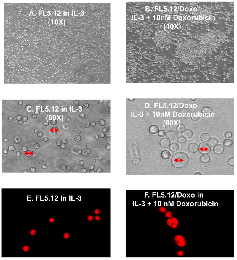
Doxorubicin resistant FL5.12 cells are larger, more blast like and some are multinucleate. The morphology of FL/5.12 and FL/Doxo cells was examined by light microscopy (Panels A & B, 10X magnification), (Panels C & D, 60X magnification). The cells were also stained with acridine orange and the nuclear morphology examined (Panels E & F).
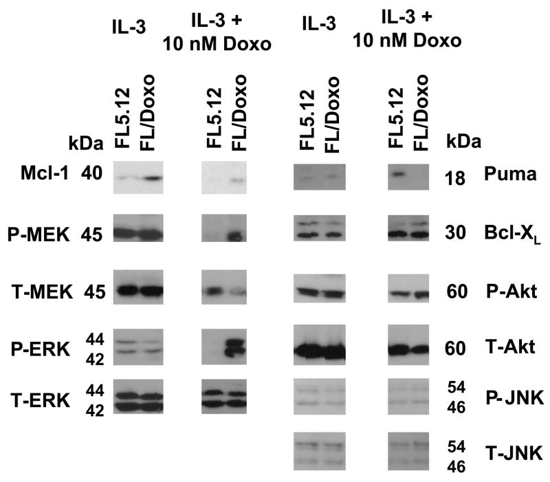
Increased activated Mcl-1, pMEK and pERK in doxorubicin resistant FL5.12 cells. FL5.12 and FL/Doxo cells were grown for 24 hours in medium containing IL-3 or IL-3 + 10 nM doxorubicin and then western blot analysis was performed with the indicated antibodies.
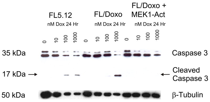
Decreased caspase 3 cleavage in doxorubicin resistant cells. The extent of cleavage of Caspase 3 was determined in doxorubicin sensitive FL5.12 cells and doxorubicin resistant FL/Doxo and FL/Doxo+MEK1-Act. The cells were incubated in the indicated concentrations of doxorubicin for 24 hours and then protein lysates isolated.
Isolation of doxorubicin resistant FL/ΔAkt:ER*+ΔRaf-1:AR cells. Limiting dilution analysis was performed in the presence of different concentrations of doxorubicin on FL/ΔAkt:ER+ΔRaf-1:AR cells. These results were obtained when the cells were cultured in medium containing 4HT + testosterone. Additional limiting dilution analyses indicated that neither 4HT nor testosterone were sufficient by themselves to result in the isolation of drug resistant cells which could be expanded into larger cultures. A dotted line is indicated at 37% of wells negative for growth from which the cloning efficiency can be estimated. These experiments have been performed 5 times and averaged together. Limiting dilution analysis with FL/ΔAkt:ER+ΔRaf-1:AR clone 1 is presented in Panel B, similar results were observed with 2 other FL/ΔAkt:ER+ΔRaf-1:AR clones.
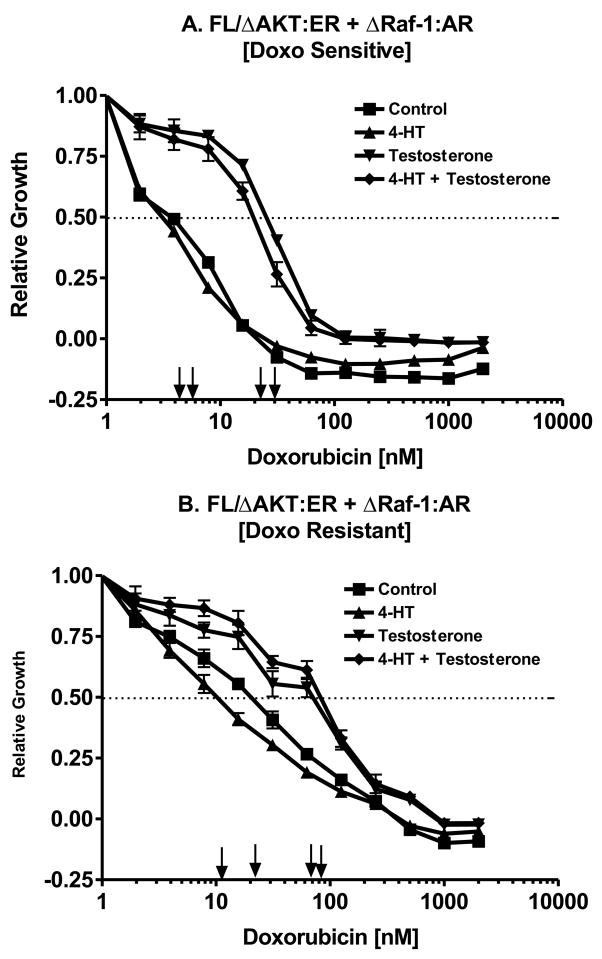
Dominant role of Raf in driving drug resistance. The effects of Raf activation by testosterone and Akt activation by 4HT on the doxorubicin IC50 of non selected and doxorubicin selected FL/ΔAkt:ER+ΔRaf-1:AR cells were examined by MTT analysis in 96 well plates. Activation of Raf increased the IC50 in both the non selected and doxorubicin selected cells.
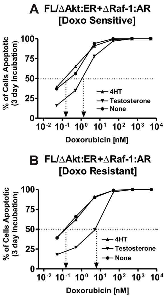
Dominant role of Raf in preventing apoptosis. The effects of Raf activation by testosterone and Akt activation by 4HT on the prevention of apoptosis induced by doxorubicin was determined in non selected and doxorubicin selected FL/ΔAkt:ER+ΔRaf-1:AR cells by the annexin V/PI technique. The extent of apoptosis was determined after incubation of the cells for 3 days in the different concentrations of doxorubicin. Cells were cultured with medium supplemented with 4HT, testosterone (test) on no supplement. Activation of Raf was dominant in the suppression of apoptosis.
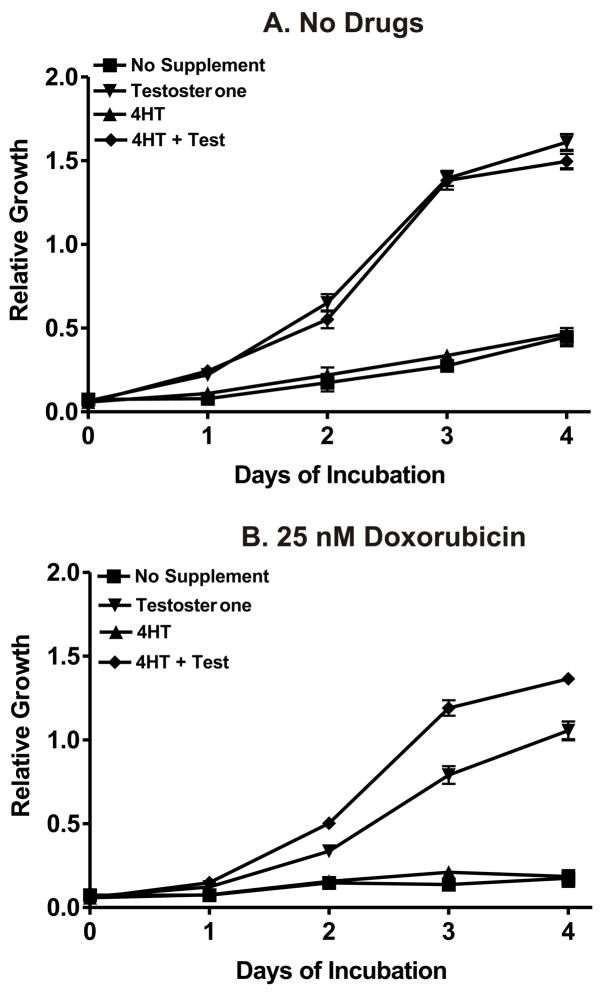
Requirement of Raf and Akt in drug resistant growth. The effects of Akt activation by 4HT, Raf activation by testosterone and their co-activation in doxorubicin selected FL/ΔAkt:ER+ΔRaf-1:AR cells was determined by MTT analysis in the presence and absence of 25 nM doxorubicin. Activation of Akt was not necessary for growth in 100 μL cultures over a 4 day period in the absence of drugs. In contrast, activation of Akt enhanced the proliferation of the cells when they were cultured in the presence of testosterone and 25 nM doxorubicin. Similar results were observed with paclitaxel and daunorubicin.
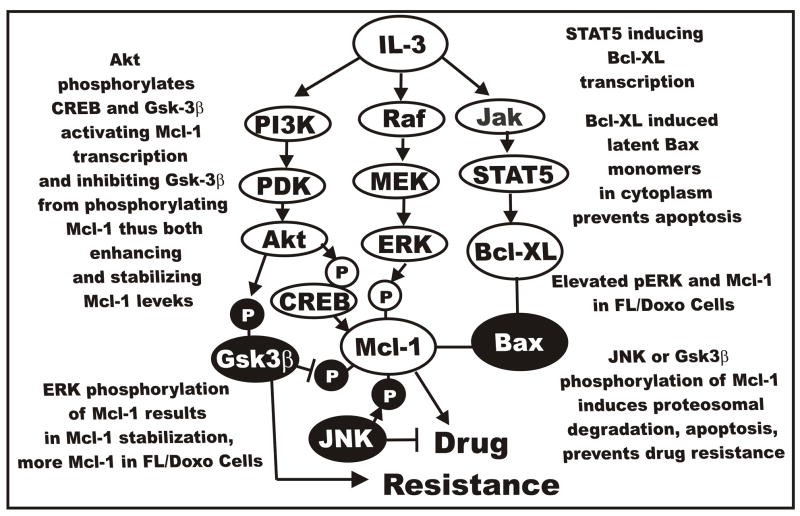
Cytokine mediated signal transduction pathways and drug resistance. Cytokines such as IL-3 can induce multiple signal transduction pathways which can effect the expression of apoptotic molecules by transcriptional and post-transcriptional mechanisms. Elevated ERK in FL/Doxo cells may phosphorylate Mcl-1 which results in its stabilization. This may result in prolonged binding to Bax, prevent activation of Bax, contribute to the prevention of apoptosis and lead to drug resistance.
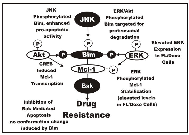
Effects of Raf>MEK>ERK and PI3K>Akt and JNK pathways on Bim phosphorylation and the induction of drug resistance. All three of these pathways can phosphorylate Bim on different residues which affect its activity and interactions with Mcl-1 and Bax and Bak. Phosphorylation events mediated by Raf>MEK>ERK and PI3K>Akt result in the prevention of Bax and Bak activation and lead to Bim being targeted to the proteosome ubiquitination and degradation. In contrast phosphorylation of Bim by JNK results in its dissociation of Bim:Mcl-1 heterodimers, Mcl-1 is targeted to the proteosome, ubiquitination, and degradation and Bim mediated activation of Bax and Bak.
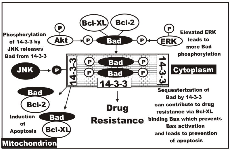
Effects of Raf>MEK>ERK and PI3K>Akt and JNK pathways on bad phosphorylation and the induction of drug resistance. All three of these pathways can phosphorylate Bad on different residues which affect its activity and interactions with Bcl-2 and Bcl-XL. Phosphorylation events mediated by Raf>MEK>ERK and PI3K>Akt result in Bad being associated with 14-3-3 proteins and translocation from the mitochondrion to the cytoplasm. Bcl-2 and Bcl-XL remain associated with Bax and Bak which prevent their activation and lead to suppression of apoptosis. In contrast phosphorylation of Bad by JNK results in its dissociation with 14-3-3 proteins and Bad localizes to the mitochondrion and binds Bcl-2 and Bcl-XL. Bax and Bak are then able to induce apoptosis.
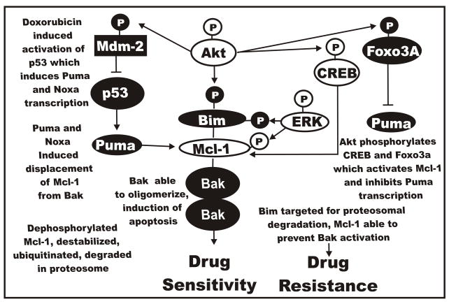
Effects of Raf>MEK>ERK, PI3K>Akt and p53 pathways on noxa and puma and the induction of drug resistance. p53 can induce the BH3 only containing Noxa and Puma proteins which interact with Mcl-1 and other anti-apoptotic proteins. When Mcl-1 is associated with Noxa and Puma that prevents their ability to interact with Bax and Bak. Increased expression of ERK in FL/Doxo cells may result in increased Mcl-1 levels which prevent Noxa and Puma abilities to activate Bax and Bak.
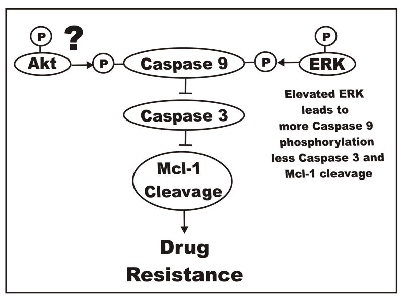
Effects of Raf>MEK>ERK and PI3K>Akt pathways on caspase 9 phosphorylation and the induction of drug resistance. The Raf>MEK>ERK pathway phosphorylates caspase 9 which prevents activation of caspase 3. The ability of Akt to phosphorylate caspase 9 is controversial as the Akt consensus phosphorylation site is present in mouse but not human caspase 9. Increased phosphorylation of caspase 9 by ERK in FL/Doxo cells could result in less caspase 9 activation, less caspase 3 activation and less Mcl-1 cleavage which could result in the prevention of apoptosis and contribute to drug resistance.
Similar articles
-
Roles of the Ras/Raf/MEK/ERK pathway in leukemia therapy.Leukemia. 2011 Jul;25(7):1080-94. doi: 10.1038/leu.2011.66. Epub 2011 Apr 15. Leukemia. 2011. PMID: 21494257 Review.
-
Dual Inhibition of PI3K-AKT-mTOR- and RAF-MEK-ERK-signaling is synergistic in cholangiocarcinoma and reverses acquired resistance to MEK-inhibitors.Invest New Drugs. 2014 Dec;32(6):1144-54. doi: 10.1007/s10637-014-0149-7. Epub 2014 Aug 26. Invest New Drugs. 2014. PMID: 25152244
-
Involvement of p53 and Raf/MEK/ERK pathways in hematopoietic drug resistance.Leukemia. 2008 Nov;22(11):2080-90. doi: 10.1038/leu.2008.207. Epub 2008 Aug 7. Leukemia. 2008. PMID: 18685611
-
Ras/Raf/MEK/ERK and PI3K/PTEN/Akt/mTOR inhibitors: rationale and importance to inhibiting these pathways in human health.Oncotarget. 2011 Mar;2(3):135-64. doi: 10.18632/oncotarget.240. Oncotarget. 2011. PMID: 21411864 Free PMC article. Review.
-
Ras/Raf/MEK/ERK and PI3K/PTEN/Akt/mTOR cascade inhibitors: how mutations can result in therapy resistance and how to overcome resistance.Oncotarget. 2012 Oct;3(10):1068-111. doi: 10.18632/oncotarget.659. Oncotarget. 2012. PMID: 23085539 Free PMC article. Review.
Cited by
-
OncomiRdbB: a comprehensive database of microRNAs and their targets in breast cancer.BMC Bioinformatics. 2014 Jan 15;15:15. doi: 10.1186/1471-2105-15-15. BMC Bioinformatics. 2014. PMID: 24428888 Free PMC article.
-
Diaporine Potentiates the Anticancer Effects of Oxaliplatin and Doxorubicin on Liver Cancer Cells.J Pers Med. 2022 Aug 16;12(8):1318. doi: 10.3390/jpm12081318. J Pers Med. 2022. PMID: 36013267 Free PMC article.
-
Targeting the PI3K/AKT/mTOR and RAF/MEK/ERK pathways for cancer therapy.Mol Biomed. 2022 Dec 21;3(1):47. doi: 10.1186/s43556-022-00110-2. Mol Biomed. 2022. PMID: 36539659 Free PMC article. Review.
-
Endothelial cell-derived interleukin-6 regulates tumor growth.BMC Cancer. 2014 Feb 17;14:99. doi: 10.1186/1471-2407-14-99. BMC Cancer. 2014. PMID: 24533454 Free PMC article.
-
Prostate cancer cells hyper-activate CXCR6 signaling by cleaving CXCL16 to overcome effect of docetaxel.Cancer Lett. 2019 Jul 10;454:1-13. doi: 10.1016/j.canlet.2019.04.001. Epub 2019 Apr 8. Cancer Lett. 2019. PMID: 30974114 Free PMC article.
References
-
- Abe J, Kusuhara M, Ulevitch RJ, Berk BC, Lee JD. Big mitogen-activated protein kinase 1 (BMK1) is a redox-sensitive kinase. J Biol Chem. 1996;271:16586–90. - PubMed
-
- Adachi T, Kar S, Wang M, Carr BI. Transient and sustained ERK phosphorylation and nuclear translocation in growth control. J Cell Physiol. 2002;192:151–59. - PubMed
-
- Aggerholm A, Gronbaek K, Guldberg P, Hokland P. Mutational analysis of the tumour suppressor gene MMAC1/PTEN in malignant myeloid disorders. Eur J Haematol. 2000;65:109–13. - PubMed
-
- Allan LA, Morrice N, Brady S, Magee G, Pathak S, Clarke PR. Inhibition of caspase-9 by phosphorylation at Thr125 by ERK MAP kinase. Nature Cell Biol. 2003;5:647–54. - PubMed
Publication types
MeSH terms
Substances
Grants and funding
LinkOut - more resources
Full Text Sources
Research Materials
Miscellaneous
