

Mammary glands and feathers: comparing two skin appendages which help define novel classes during vertebrate evolution
- PMID: 17382566
- PMCID: PMC4382004
- DOI: 10.1016/j.semcdb.2007.02.005
Mammary glands and feathers: comparing two skin appendages which help define novel classes during vertebrate evolution
Abstract
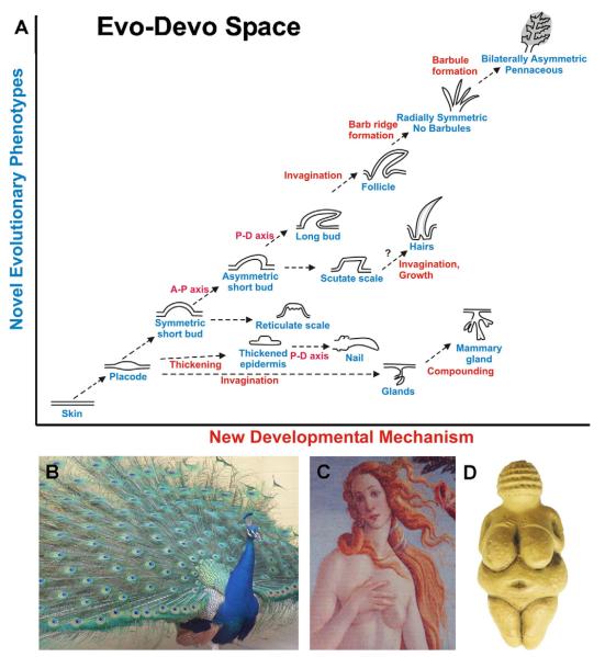
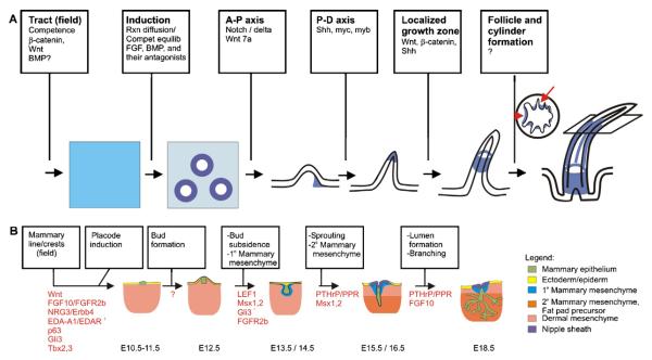
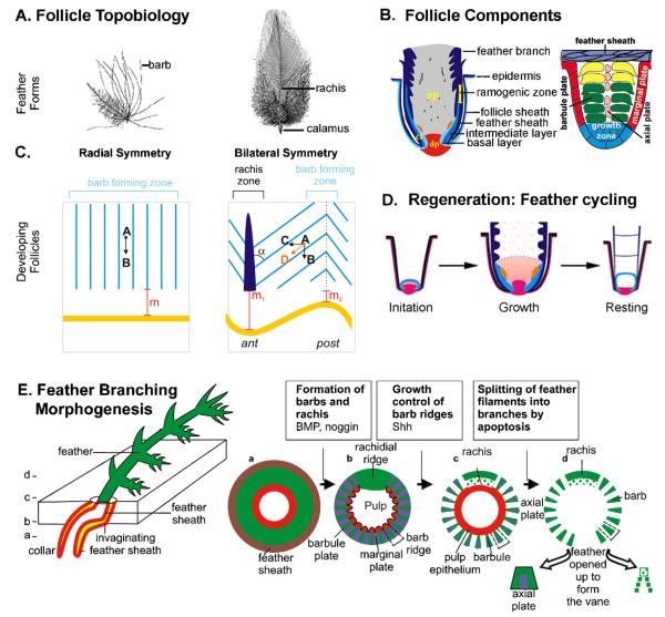
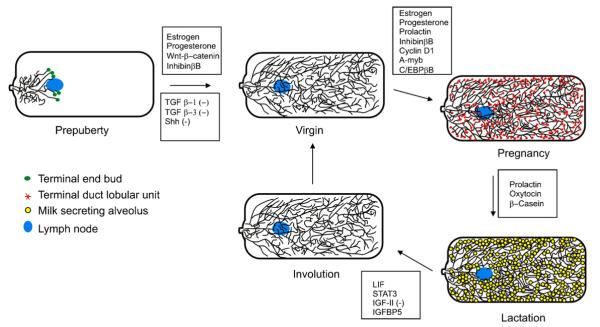
It may appear counter-intuitive to compare feathers and mammary glands. However, through this Evo-Devo analysis, we appreciate how species interact with the environment, requiring different ectodermal organs. Novel ectodermal organs help define evolutionary directions, leading to new organism classes as exemplified by feathers for Aves and mammary glands for Mammals. Here, we review their structure, function, morphogenesis and regenerative cycling. Interestingly, both organs undergo extensive branching for different reasons; feather branching is driven by mechanical advantage while mammary glands nourish young. Besides natural selection, both are regulated by sex hormones and acquired a secondary function for attracting mates, contributing to sexual selection.
Figures
Similar articles
-
Evo-Devo of amniote integuments and appendages.Int J Dev Biol. 2004;48(2-3):249-70. doi: 10.1387/ijdb.041825pw. Int J Dev Biol. 2004. PMID: 15272390 Free PMC article. Review.
-
Development and evolution of the amniote integument: current landscape and future horizon.J Exp Zool B Mol Dev Evol. 2003 Aug 15;298(1):1-11. doi: 10.1002/jez.b.23. J Exp Zool B Mol Dev Evol. 2003. PMID: 12949766 Free PMC article.
-
Avian skin development and the evolutionary origin of feathers.J Exp Zool B Mol Dev Evol. 2003 Aug 15;298(1):57-72. doi: 10.1002/jez.b.26. J Exp Zool B Mol Dev Evol. 2003. PMID: 12949769 Review.
-
SnapShot: Branching Morphogenesis.Cell. 2014 Aug 28;158(5):1212-1212.e1. doi: 10.1016/j.cell.2014.08.019. Cell. 2014. PMID: 25171418 Free PMC article.
-
Organizational principles of integumentary organs: Maximizing variations for effective adaptation.Dev Biol. 2025 Jun;522:171-195. doi: 10.1016/j.ydbio.2025.03.011. Epub 2025 Mar 18. Dev Biol. 2025. PMID: 40113027 Review.
Cited by
-
Evo-devo of the mammary gland.J Mammary Gland Biol Neoplasia. 2013 Jun;18(2):105-20. doi: 10.1007/s10911-013-9290-8. Epub 2013 May 17. J Mammary Gland Biol Neoplasia. 2013. PMID: 23681303 Review.
-
A shark-inspired general model of tooth morphogenesis unveils developmental asymmetries in phenotype transitions.Proc Natl Acad Sci U S A. 2023 Apr 11;120(15):e2216959120. doi: 10.1073/pnas.2216959120. Epub 2023 Apr 7. Proc Natl Acad Sci U S A. 2023. PMID: 37027430 Free PMC article.
-
The Hair Follicle: An Underutilized Source of Cells and Materials for Regenerative Medicine.ACS Biomater Sci Eng. 2018 Apr 9;4(4):1193-1207. doi: 10.1021/acsbiomaterials.7b00072. Epub 2017 Mar 21. ACS Biomater Sci Eng. 2018. PMID: 29682604 Free PMC article.
-
De Novo Assembly and Comparative Transcriptome Profiling of Anser anser and Anser cygnoides Geese Species' Embryonic Skin Feather Follicles.Genes (Basel). 2019 May 8;10(5):351. doi: 10.3390/genes10050351. Genes (Basel). 2019. PMID: 31072014 Free PMC article.
-
Development, regeneration, and evolution of feathers.Annu Rev Anim Biosci. 2015;3:169-95. doi: 10.1146/annurev-animal-022513-114127. Epub 2014 Nov 3. Annu Rev Anim Biosci. 2015. PMID: 25387232 Free PMC article. Review.
References
-
- Chuong CM, editor. Molecular basis of epithelial appendage morphogenesis. Landes Bioscience; Austin: 1998. pp. 3–14.
-
- Oftedal OT. The mammary gland and its origin during synapsid evolution. J Mammary Gland Biol Neoplasia. 2002;7:225–52. - PubMed
Publication types
MeSH terms
Grants and funding
LinkOut - more resources
Full Text Sources
