. 2007 Mar 23;128(6):1105-18.
doi: 10.1016/j.cell.2007.01.038.

AU-rich-element-mediated upregulation of translation by FXR1 and Argonaute 2

Affiliations

AU-rich-element-mediated upregulation of translation by FXR1 and Argonaute 2

Shobha Vasudevan et al. Cell. .

Abstract

AU-rich elements (AREs), present in mRNA 3'-UTRs, are potent posttranscriptional regulatory signals that can rapidly effect changes in mRNA stability and translation, thereby dramatically altering gene expression with clinical and developmental consequences. In human cell lines, the TNFalpha ARE enhances translation relative to mRNA levels upon serum starvation, which induces cell-cycle arrest. An in vivo crosslinking-coupled affinity purification method was developed to isolate ARE-associated complexes from activated versus basal translation conditions. We surprisingly found two microRNP-related proteins, fragile-X-mental-retardation-related protein 1 (FXR1) and Argonaute 2 (AGO2), that associate with the ARE exclusively during translation activation. Through tethering and shRNA-knockdown experiments, we provide direct evidence for the translation activation function of both FXR1 and AGO2 and demonstrate their interdependence for upregulation. This novel cell-growth-dependent translation activation role for FXR1 and AGO2 allows new insights into ARE-mediated signaling and connects two important posttranscriptional regulatory systems in an unexpected way.


Figures
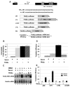
Figure 1
Figure 1. In Vivo Assay for ARE-Regulated Translation
(A) TNFα mRNA with conserved regions, including a splicing regulator (APRE; Osman et al., 1999), the ARE (AU), and a constitutive decay element (CDE; Stoecklin et al., 2003) are shown. Firefly luciferase reporter constructs with the 34 nt TNFα ARE (ARE), the 795 nt TNFα 3′-UTR (UTR), the 34 nt mutant ARE (mt ARE), or a 34 nt vector sequence (CTRL) were cotransfected with a Renilla luciferase reporter (REN). Mutations in mt ARE and regions protected by the probes used in RNase protection assays (RPAs) are underlined. (B) HEK293 cells were transfected with the ARE and REN reporters and 18 hr later were switched to serum-containing (+) or serum-lacking (−) medium containing either TPA/Io (see Experimental Procedures) or the DMSO solvent. Eighteen hours later, firefly and Renilla luciferase activities were assayed. The values in panels (B), (D), and (E) are averages from at least three transfections ± SD. (C) Northern blots show ARE- and REN-reporter levels in ± serum, with TPA/Io, Io alone, and/or the solvent DMSO as indicated. Table S2 shows the RNA values. (D) Luciferase values (B) were normalized to the mRNA levels (C) to obtain translation efficiencies (defined in Figure 1A; see Figure S1 and Tables S1 and S2). Since similar changes upon serum starvation without TPA/Io were observed without normalizing for RNA levels (B), normalization does not artificially produce an apparent increase in translation. (E) Comparison of translation efficiencies of the ARE and UTR reporters relative to the control reporters, CTRL, and mt ARE in response to the presence or absence of serum without DMSO is shown.
Figure 2
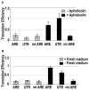
Figure 2. Cell-Cycle Arrest Increases Translation Mediated by the TNFα ARE
(A) Eighteen hours after transfection as in Figure 1, HEK293 cells were treated with aphidicolin dissolved in DMSO or with DMSO alone for 36 hr before measuring luciferase activities and mRNA levels by RPA. The values in panels (A) and (B) are averages from at least three transfections ± SD. (B) After transfection, cells were grown for 66 hr to saturation. Then, one set was replated in fresh media; six hours later, luciferase and mRNA levels were assessed. Cell-cycle arrest was validated by western blotting for Ki67 (Figure S3 and Table S3).
Figure 3
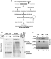
Figure 3. Purification of mRNP Complexes
(A) After in vivo formaldehyde crosslinking, the S1-tagged ARE construct was purified as described in Experimental Procedures. An S1-tagged CTRL construct, an untagged ARE reporter, and uncrosslinked controls were examined in parallel. (B) RPA demonstrates enrichment of the ARE and CTRL S1-tagged RNAs (lanes 9–12) in the biotin eluates compared to the nontagged ARE reporter (lanes 7 and 8) and the cotransfected REN internal control. Unmarked bands are heat-induced degradation products of the firefly ARE reporter. –Crosslink (uncrosslinked extract) and –Extract (no sample) served as RPA controls. (C) Proteins present in crosslinked eluates from the S1-tagged UTR, ARE, and CTRL constructs in the basal translation serum-grown conditions (lanes 1, 3, and 5) were compared to those in the activated serum-starved conditions (lanes 2, 4, and 6) by western blot analysis. α-UPF3 served as control. The unlabeled lane between lanes 2 and 3 was from a TPA-treated UTR sample.
Figure 4
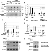
Figure 4. FXR1 Is Required for TNFα-ARE-Mediated Translation Upregulation in Response to Serum Starvation
(A) Knockdown of FXR1 by shRNA FA4, predicted to target FXR1 iso-a (see Figure S5A), is shown. Western blotting (α-FXR1; described in Jin et al., 2004) reveals FXR1 in the soluble fraction only in serum-starved conditions (upper panel, lanes 1–4, 6, and 8) compared to sonicated total extracts from the same cells (lower panel). Two-fold titrations of the serum-starved extract (lanes 1–4) are compared with extracts (the same amount as in lane 4) from cells grown with serum (+ lanes) or without serum (− lanes) that were either untreated (lanes 5 and 6) or exposed to either shRNA FA3 (lanes 7 and 8) or shRNA FA4 (lanes 9 and 10). NS is a nonspecific band or breakdown product recognized by the anti-FXR1 polyclonal antibody. (B) Knockdown of FXR1 by shRNA FA4 leads to loss of translation activation of the ARE relative to the CTRL reporter in serum-starved conditions. The values in panels (B), (C), and (E) are averages from at least three transfections ± SD. (C) Exogenous expression of the isoform, FXR1 iso-a (the most abundant isoform in our cells), increases the translation efficiency of the ARE and UTR reporters but not of the mt ARE in serum-grown conditions. Western blotting (below) demonstrates that exogenous expression of iso-a generates FXR1 in the soluble fraction of serum-grown cells (+ lanes). The band at 50 kDa is nonspecific and is described in (A). Anti-tubulin provided a loading control. (D) The 5B Box tethering site of 343 nt (Baron-Benhamou et al., 2004) replaces the ARE in the firefly ARE reporter. (E) Flag-tagged FXR1 iso-a tethered by the λN peptide to the 5B Box reporter activates translation in both serum conditions compared to the CTRL reporter or the empty λN vector. (F) Western blotting as in (C) reveals similar expression of the λN-peptide- and Flag-tagged FXR1 iso-a in soluble extracts from serum-grown and -starved cells. The middle panel discriminates the Flag-tagged and endogenous FXR1 isoforms. Comparable results were obtained with cells grown to saturation (quiescence).
Figure 5
Figure 5. AGO2 Is Present in the ARE Translation Activation Complex
(A) FXR1 interacts with AGO2 in serum-grown and -starved HEK293 cells. In vivo formaldehyde crosslinking of cells that were untransfected or transfected with the λN-FXR1 iso-a-Flag vector was followed by sonicated cell-extract preparation. Immunoprecipitation of the untransfected sample (lanes 3–8) with α-FXR1 (lanes 5–8) and of the λN-FXR1 iso-a-Flag-transfected sample (lanes 1, 2, and 9–14) with either α-Flag (lanes 9–12) or α-HA (lanes 13 and 14, negative control) was followed by stringent washing. Western blotting (α-AGO2 antibody; Upstate) revealed AGO2 coimmunoprecipitation with α-FXR1 (lanes 5 and 6) or α-Flag (lanes 9 and 10) from crosslinked samples only (lanes 7 and 8 for α-FXR1 IP and lanes 11 and 12 for α-Flag IP-uncrosslinked). The absence of reporters (this Figure) or the presence of ARE, 5B Box, or control reporters did not alter the interaction (data not shown). No RNase (this Figure) or RNase-treated extracts (RNase A and RNase One, data not shown) showed similar interactions. (B) Confocal immunofluorescence imaging was performed with fixed samples of serum-grown and -starved HEK293 cells stained with α-AGO2, α-FXR1, or α-GW182. TO-PRO-3 (Invitrogen) identified DNA in the nuclei. FXR1 staining was identical using polyclonal antibody from S. Warren or monoclonal antibody (6GB10) from G. Dreyfuss. Standard controls using single antibodies and no primary antibody ensured no artifactual channel overlap/staining (data not shown). Magnified images and details are in Figures S7–S8. (C) RNPs formed on the S1-tagged ARE and CTRL reporters were isolated as in Figure 3A, which was followed by western analysis of the eluates (lanes 3–8) with α-AGO2. AGO2 appeared specifically in translation activation conditions (compare lanes 5 [serum +] and 7 [serum −]). The extracts (input) from noncrosslinked cells serve as negative controls (lanes 6 and 8), as do eluates from the S1-tagged CTRL reporter (lanes 3 and 4). (D) Anti-AGO2 immunoprecipitates the S1-tagged ARE reporter exclusively from translation activation (serum-starved) conditions (lane 4 versus lane 3). Immunoprecipitation was performed as in (A) on extracts of crosslinked cells. The immunoprecipitates (lanes 1–6) and inputs (extracts, lanes 7–12) were analyzed by RPA. Cells expressing the CTRL (lanes 1 and 2) reporter served as a negative control.
Figure 6
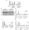
Figure 6. AGO2 Activates Translation Mediated by TNFα ARE in Response to Serum Starvation and Requires FXR1 to Function as a Translation Activator
(A) Tethering AGO2 upregulates translation in response to serum starvation. A mutant form of AGO2 (mt AGO2; Pillai et al., 2004) and the CTRL reporter (Figure 1A) without the 5B Boxes provided negative controls. FXR1 iso-a (Figure 4E) served as a positive control. The values in panels (A), (C), (D), and (E) are averages from at least three transfections ± SD. (B) Western blot done using α-AGO2 of soluble HEK293 extracts to assess knockdown by two shRNAs used together at either 0.5 μg or 1.0 μg plasmid DNA (shAGO2; see Experimental Procedures) is shown. Two extract concentrations and α-tubulin antibody provided controls. (C) Comparison of the effects of no shRNA (−), FXR1 shRNA (FA4), or AGO2 shRNA (shAGO2) on the translation efficiencies of the ARE and mt ARE reporters under serum+ and serum– conditions shows that both FXR1 and AGO2 are required for upregulation. (D) FXR1 is essential for translation upregulation mediated by tethered AGO2. AGO2 or the λN-tag vector (as a control) was tethered to the firefly reporter in control or FXR1 shRNA (FA4)-treated cells under serum-grown and -starved conditions. (E) AGO2 is essential for translation upregulation mediated by tethered FXR1. λN-tagged FXR1 iso-a or the λN-tag vector was transfected with the firefly reporter bearing 5B Boxes in control (−) or AGO2 shRNA (shAGO2)-treated cells under serum-grown and -starved conditions. Comparable results were obtained with cells grown to saturation (quiescence).
Figure 7
Figure 7. Serum Starvation Activates Translation by Relocalization of AGO2-Bound mRNPs to Polysomes
(A) Polysome profiles of extracts from (1) serum-grown or (2) serum-starved cells transfected with the TNFα ARE are shown. (B) Western analysis of fractions across the profiles using antibodies against AGO2, FXR1, and PABC1 as a control is shown. FXR1 mobility becomes heterogeneous (modifications or protein degradation) upon serum starvation. Since the lysates were clarified, the majority of FXR1 and AGO2 in +serum are removed as large complexes, including those in large foci (Figures 5B and S7–S8), prior to gradient analysis (verified by the Extract lanes marked +serum and −serum). (C) Anti-AGO2 (marked by brackets) and Y10B (arrows) immunoprecipitations were performed on the Free RNP (fractions 1–2), subunits (fractions 3–9, including monosomes), and polysomes (fractions 10–22), followed by western analyses using α-AGO2 and -FXR1 antibodies. (D) RPA for the ARE-reporter RNA was performed on the pooled fraction as in (C). (E) Western analysis of polysome profiles from serum-grown and -starved cells demonstrates a complete shift of AGO2 from polysomes (compare with B, −Serum lanes) to a single slowly sedimenting peak upon 1 mM puromycin treatment for 3 hr at 37°C. Puromycin causes an equal proportion of AGO2 to be clarified from extracts of both serum conditions.

Comment in

Similar articles

Cited by

References

    1. Andersson K, Sundler R. Signalling to translational activation of tumour necrosis factor-alpha expression in human THP-1 cells. Cytokine. 2000;12:1784–1787. - PubMed
    1. Andrei MA, Ingelfinger D, Heintzmann R, Achsel T, Rivera-Pomar R, Luhrmann R. A role for eIF4E and eIF4E-transporter in targeting mRNPs to mammalian processing bodies. RNA. 2005;11:717–727. - PMC - PubMed
    1. Bakheet T, Frevel M, Williams BR, Greer W, Khabar KS. ARED: human AU-rich element-containing mRNA database reveals an unexpectedly diverse functional repertoire of encoded proteins. Nucleic Acids Res. 2001;29:246–254. - PMC - PubMed
    1. Balkwill F. Tumor necrosis factor or tumor promoting factor? Cytokine Growth Factor Rev. 2002;13:135–141. - PubMed
    1. Baron-Benhamou J, Gehring NH, Kulozik AE, Hentze MW. Using the lambdaN peptide to tether proteins to RNAs. Methods Mol Biol. 2004;257:135–154. - PubMed

Publication types

MeSH terms