. 2007 May 15;305(2):564-76.
doi: 10.1016/j.ydbio.2007.02.037. Epub 2007 Mar 3.

E2f4 is required for normal development of the airway epithelium

Affiliations

E2f4 is required for normal development of the airway epithelium

Paul S Danielian et al. Dev Biol. .

Abstract

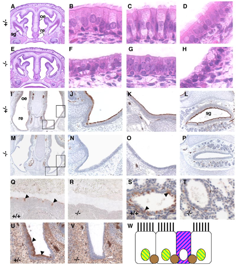
The airway epithelium is comprised of specialized cell types that play key roles in protecting the lungs from environmental insults. The cellular composition of the murine respiratory epithelium is established during development and different cell types populate specific regions along the airway. Here we show that E2f4-deficiency leads to an absence of ciliated cells from the entire airway epithelium and the epithelium of the submucosal glands in the paranasal sinuses. This defect is particularly striking in the nasal epithelium of E2f4-/- mice where ciliated cells are replaced by columnar secretory cells that produce mucin-like substances. In addition, in the proximal lung, E2f4 loss causes a reduction in Clara cell marker expression indicating that Clara cell development is also affected. These defects arise during embryogenesis and, in the nasal epithelium, appear to be independent of any changes in cell proliferation, the principal process regulated by members of the E2f family of transcription factors. We therefore conclude that E2f4 is required to determine the appropriate development of the airway epithelium. Importantly, the combination of no ciliated cells and excess mucous cells can account for the chronic rhinitis and increased susceptibility to opportunistic infections that causes the postnatal lethality of E2f4 mutant mice.


Figures

Fig. 1
Fig. 1
Mutation of E2f4 leads to a dramatic reduction in the number of ciliated cells only in the airway epithelium. The presence of motile cilia on epithelia was assessed by immunohistochemistry using an antibody against acetylated α-tubulin that is a component of motile cilia (brown stain). Nasal epithelium from a control 3 months old E2f4+/− mouse (A) is highly ciliated in contrast to that of an E2f4−/− age matched littermate (B). Cilia were not detected in the bronchioles of 2.5 months old E2f4−/− mice (D) in contrast to an age matched wild-type littermate (C). The ciliated epithelium in the fallopian tube (E,F), at 7 months, the efferent ducts of the epididymis (G,H) and ependymal lining of the brain ventricles (I,J) at 2.5 months, of E2f4−/− mice appeared similar to that of the age matched littermate controls. Sperm flagella (indicated by the *) appear to be normal in E2f4−/− mice at 2.5 months (L) in comparison with control mice (K). Original magnification ×40. Arrowheads point to ciliated cells in B and C.
Fig. 2
Fig. 2
E2f4 mutant embryos lack ciliated cells in the airway epithelium. (A–H) Hematoxylin and eosin stained sections from E2f4+/− (A–D) and E2f4 mutant (E–H) 18.5 dpc littermate embryos. Panels A and E, coronal sections (original magnification ×4) showing the respiratory epithelium (re), olfactory epithelium (oe) and paranasal sinus serous gland (sg). High power images (original magnification ×40) of the respiratory epithelium (B, F), serous gland epithelium (C, G) and nasopharynx epithelium (D, H). (I–V) Acetylated α-tubulin, a component of cilia, was visualized by immunohistochemistry (brown stain). Panels I and M, coronal sections (original magnification ×10) with rectangles showing positions of the two adjacent panels. No staining was observed in the epithelium of E2f4 mutant embryos (M–O) compared with control embryos (I–K) (original magnification ×40) at 18.5 dpc. This phenotype was also observed in the serous gland (L, P) (original magnification ×20), in the trachea (Q, R) and bronchioles (S, T) at 18.5 dpc and in the nasal respiratory epithelium at 16.5 dpc (U, V) (original magnification ×40). Control genotypes are indicated (Q, S and U), E2f4−/− (R, T and V). Note that acetylated α-tubulin is also detected in neurons. Arrowheads point to ciliated cells in Q, S and U. W, Schematic of wild-type nasal respiratory epithelium with cell specific markers. Ciliated cells express Foxj1 (yellow) and Foxa1 (green). Goblet cell mucins stain with PAS (magenta) and Alcian Blue (blue). Basal cells express p63 (brown).
Fig. 3
Fig. 3
Analyses of proliferation in the E2f4 mutant epithelium. Representative adjacent coronal sections from 16.5 dpc wild-type (A,B, E,F and I,J) or E2f4 mutant littermate embryos (C, D, G,H, and K,L) analyzed by immunohistochemistry (brown stain) to assess the incorporation of BrdU (A–D), expression of Ki67 (E–H) and PCNA (I–L). Dorsal is to the left, original magnification ×40. The line marks the junction between the olfactory epithelium (oe) and respiratory epithelium (re). The outlined rectangle marks the area displayed in the adjacent panel. Slightly higher intensity PCNA staining was detected throughout E2f4 −/− embryos. M, Quantification of BrdU and Ki67 staining is shown at the embryonic stages indicated. A minimum of four pairs of wild-type and E2f4+/− (Ct) or E2f4 −/− (−/−) littermate embryos were analyzed at each stage. The means with standard deviation error bars are shown. Performance of the paired Student’s t-test indicated that none of the pairs was significantly different (all p values >0.05) apart from the 18.5 dpc BrdU time point (p = 0.025).
Fig. 4
Fig. 4. Cells comprising the E2f4 mutant nasal respiratory epithelium contain mucins
Adjacent matched sections from wild-type or E2f4 +/− (A–C, G–I and M–O) or E2f4−/−embryos (D–F, J–L and P–R) at 18.5 dpc were subjected to the PAS reaction (A–F) that stains neutral mucins (pink stain), Alcian Blue pH2.5 stain (G–L) that stains acidic mucins and immunohistochemistry to detect MUC5AC, brown stain (M–R). Panels A,D,G,J,M and P lower power (original magnification ×20) images of coronal sections containing respiratory epithelium. Panels B,E,H,K,N and Q higher power images (original magnification ×40) of the area encompassed by the rectangle in the adjacent panel. Panels C,F,I,L,O and R sections of the nasopharynx from the same embryo as in the adjacent panels. In the control samples (wild-type or E2f4 +/−) only goblet cells stain for mucins whilst most of the cells within the E2f4 mutant epithelium stain for a mucin marker. PAS also stains bone and Alcian Blue also stains cartilage.
Fig. 5
Fig. 5
Immunohistochemical analyses of epithelium specific markers. Matched sections from 18.5 dpc wild-type or E2f4+/− as indicated (A,C) or E2f4−/− littermate embryos (B,D) were stained for cytokeratin 8 (A,B), a general marker for epithelial cells, and Foxa1 (C,D) a specific marker for proximal respiratory epithelial cells (brown stain). Note that Foxa1 is not expressed in goblet cells (stained with Alcian Blue) in the control samples. Expression of Foxj1 at 18.5 dpc (E,F) and 15.5 dpc (G,H) a marker of ciliated epithelial cells was absent from the nuclei of the E2f4−/− embryo epithelium (F,H) in contrast to the control samples (E,G), brown stain. Basal cells were detected by analyzing the expression of p63 at 18.5 dpc (I,J) and 16.5 dpc (K,L). In controls (I,K), in addition to the basal cells, nuclei in the columnar epithelial cells stained for p63 protein (brown stain) whereas this staining was not detected in the epithelium of E2f4−/− embryos (J,L). E2f4 was detected in many of the nuclei of control (wild-type or E2f4+/−) embryo epithelial cells at 18.5 dpc (M) and 15.5 dpc (O), brown stain. Staining was absent from matched sections of E2f4−/− embryos (N,P). E2f4 was also detected in most nuclei in the lung at 18.5 dpc including those in the bronchiole epithelium (Q) and staining was absent from the E2f4−/− embryo lung (R). At 18.5 dpc CC10, a marker of Clara cells, was observed in the bronchioles of E2f4−/− embryos (T) at a reduced level in comparison with E2f4+/− embryos (S). T1α, a marker for type I alveolar cells was expressed on the apical membrane at similar levels in wild-type (U) and E2f4−/− embryos (V) at 18.5 dpc. Cells staining for SP-C (black stain), a marker for type II alveolar cells are detected at similar frequency in E2f4+/− (W) and E2f4−/− embryos (X) at 18.5 dpc. Sections in panels C–F,I,J were additionally stained using the Alcian Blue pH2.5 method to identify cells containing acidic mucins. Original magnification ×40.
Fig. 6
Fig. 6. Transmission electron microscopy analyses of E2f4−/− nasal epithelium
Control (wild-type or E2f4+/−) embryo epithelium contained ciliated columnar cells interspersed with goblet cells (arrowhead), * denotes cilia and the arrow points to microvilli (A). Epithelium of E2f4−/− embryos lacks cilia and is comprised of cells resembling columnar secretory cells that contained high amounts of rough endoplasmic reticulum and secretory vesicles (arrowheads) distributed apically (B) and throughout the cell, arrow indicates microvilli (C). D, higher power image of the area delineated by the box in C showing dilated rough endoplasmic reticulum. Scale bar in A, B and C 2μm, in D 500nm.

References

    1. Apostolova MD, et al. Active nuclear import and export pathways regulate E2F-5 subcellular localization. J Biol Chem. 2002;277:34471–9. - PubMed
    1. Attwooll C, et al. The E2F family: specific functions and overlapping interests. Embo J. 2004;23:4709–16. - PMC - PubMed
    1. Banizs B, et al. Dysfunctional cilia lead to altered ependyma and choroid plexus function, and result in the formation of hydrocephalus. Development. 2005;132:5329–39. - PubMed
    1. Besnard V, et al. Immunohistochemical localization of Foxa1 and Foxa2 in mouse embryos and adult tissues. Gene Expr Patterns. 2004;5:193–208. - PubMed
    1. Blatt EN, et al. Forkhead transcription factor HFH-4 expression is temporally related to ciliogenesis. Am J Respir Cell Mol Biol. 1999;21:168–76. - PubMed

Publication types

MeSH terms

Substances