. 2007 May 11;368(4):966-81.
doi: 10.1016/j.jmb.2007.02.046. Epub 2007 Feb 22.

Genome sequence, structural proteins, and capsid organization of the cyanophage Syn5: a "horned" bacteriophage of marine synechococcus

Affiliations

Genome sequence, structural proteins, and capsid organization of the cyanophage Syn5: a "horned" bacteriophage of marine synechococcus

Welkin H Pope et al. J Mol Biol. .

Abstract

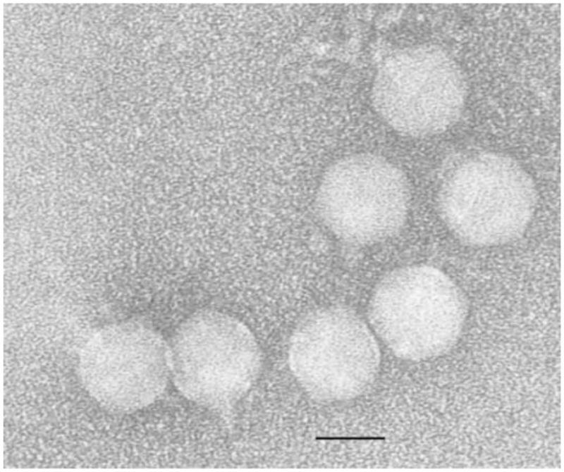
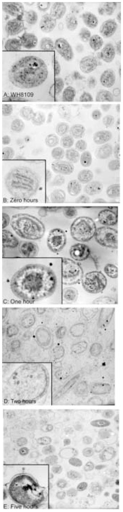
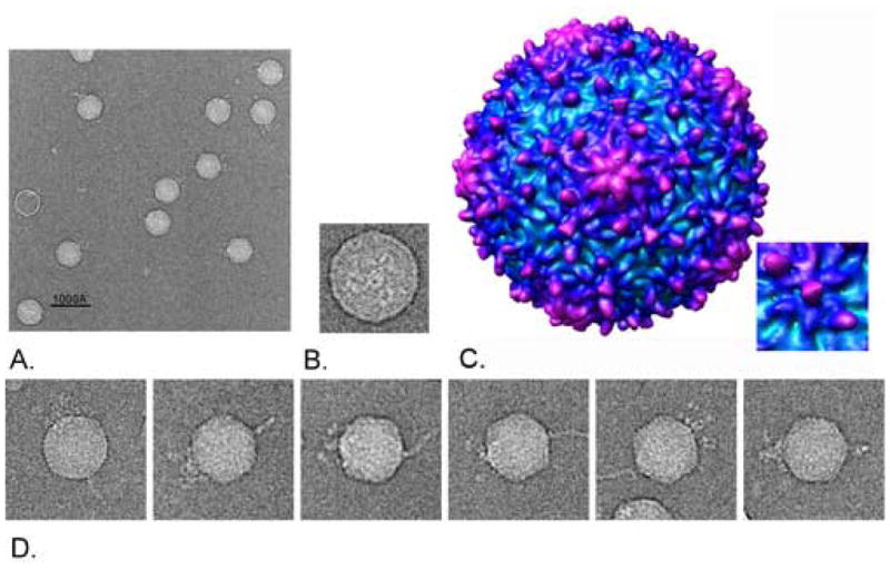
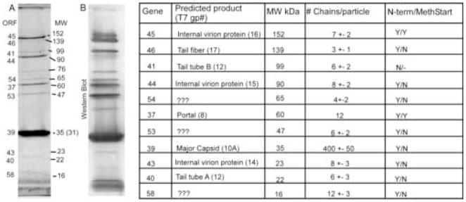
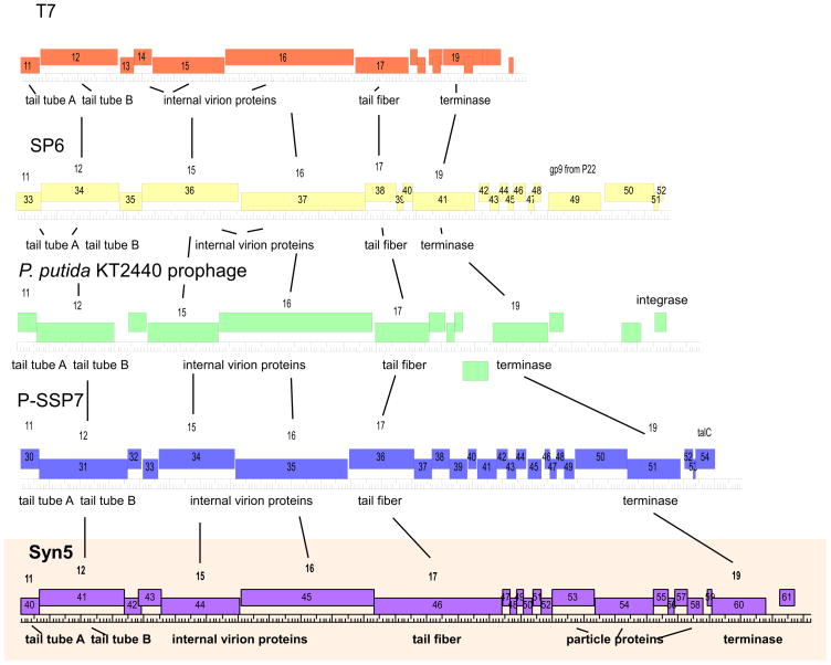
Marine Synechococcus spp and marine Prochlorococcus spp are numerically dominant photoautotrophs in the open oceans and contributors to the global carbon cycle. Syn5 is a short-tailed cyanophage isolated from the Sargasso Sea on Synechococcus strain WH8109. Syn5 has been grown in WH8109 to high titer in the laboratory and purified and concentrated retaining infectivity. Genome sequencing and annotation of Syn5 revealed that the linear genome is 46,214 bp with a 237 bp terminal direct repeat. Sixty-one open reading frames (ORFs) were identified. Based on genomic organization and sequence similarity to known protein sequences within GenBank, Syn5 shares features with T7-like phages. The presence of a putative integrase suggests access to a temperate life cycle. Assignment of 11 ORFs to structural proteins found within the phage virion was confirmed by mass-spectrometry and N-terminal sequencing. Eight of these identified structural proteins exhibited amino acid sequence similarity to enteric phage proteins. The remaining three virion proteins did not resemble any known phage sequences in GenBank as of August 2006. Cryo-electron micrographs of purified Syn5 virions revealed that the capsid has a single "horn", a novel fibrous structure protruding from the opposing end of the capsid from the tail of the virion. The tail appendage displayed an apparent 3-fold rather than 6-fold symmetry. An 18 A resolution icosahedral reconstruction of the capsid revealed a T=7 lattice, but with an unusual pattern of surface knobs. This phage/host system should allow detailed investigation of the physiology and biochemistry of phage propagation in marine photosynthetic bacteria.


Figures

Figure 1
Figure 1
Electron Micrograph of Purified Syn5 Virions. Purified particles were placed on copper mesh grids and stained with 2% uranyl acetate. Particles exhibited the same morphology as originally observed by Waterbury and Valois; with an isometric icosahedral capsid approximately 60nm in diameter, and a short tail approximately 25nm in length.
Figure 2
Figure 2
Cryoelectron micrographs of purified Syn5 particles and Icosahedral 18Å Reconstruction of the Capsid. Syn5 particles were flash-frozen and examined by cryoelectron microscopy. A) Syn5 virions. The extended horn is seen directly opposite the three-pronged tail on the isometric capsid. Scale bar = 100nm B) End view of the Syn5 particle. The tail tube is surrounded by tail proteins with threefold symmetry. C) 18Å Capsid Reconstruction with imposed icosahedral symmetry. The 18Å icosahedral reconstruction of the capsid is shown radially colored from cyan to purple with increasing radius. The map is in the T=7L form, but the hand of the capsid has yet to be resolved. D) Gallery of single Syn5 particles with horn structures.
Figure 3
Figure 3
Thin-sections of Synechococcus WH8109 infected with Cyanophage Syn5. Time course of WH8109 cells infected with Syn5 in late exponential phase. A) Uninfected cells. B) Time Zero: phage were mixed with cells and immediately fixed with glutaraldehyde. C–E) Phage and cells were incubated for one, two, and five hours prior to fixation.
Figure 4
Figure 4
Map of the Syn5 Genome. The Syn5 genome is 46,214bp long with a 237bp terminal repeat. 61 genes are labeled. Genes identified with BLASTp as having similarity to known phage DNA replication genes are in blue, to structural proteins are in red, and to non-structural late genes in green. Unidentified genes are in magenta. Also labeled are the putative integrase (int), ribonucleotide reductase (nrt), thioredoxin (trd), and thymidylate synthase (thy) genes.
Figure 5
Figure 5
SDS-polyacrylamide gel and Western Blot of Syn5 virion proteins. Purified Syn5 particles were electrophoresed through 10% polyacrylamide containing SDS. A) Polyacrylamide gel stained with Coomassie Blue. 12 protein bands are visible and are marked with: the encoding gene number as determined by tandem mass-spectrometry, the approximate molecular weights in kDa as determined by relative mobility, polypeptide chain copy number rounded to the nearest whole number followed by the equivalent protein’s copy number in T7 in parentheses, confirmation of mass-spectrometric identification by N-terminal sequencing (Y for yes, N for no) and presence of N-terminal methionine on the protein (Y for yes, N for no). B) Western blot. After electrophoresis, proteins were blotted to PVDF membrane and probed with polyclonal antiSyn5 rabbit sera. The bands were then visualized using the ECF anti-rabbit kit (Amersham)
Figure 6
Figure 6
Structural arms of short-tailed phage genomes. Comparison of structural arms of five T7-like phages. Genes are represented by rectangles, and labeled both according to T7 nomenclature (above) and the number at which they occur when counting genes from the left end of the genome to the right end of the genome (inside). Each full length hatch mark on the ruler = 1kb. The synteny and conservation of length of the T7-like genes at the right end of the genome is readily apparent, as is the plasticity at the extreme right end genomes with regards to the inclusion of an additional gene module. Figure generated by DNA Master.

Similar articles

Cited by

References

    1. Partensky F, Hess WR, Vaulot D. Prochlorococcus, a marine photosynthetic prokaryote of global significance. Microbiol Mol Biol Rev. 1999;63:106–127. - PMC - PubMed
    1. Waterbury JB, Watson SW, Valois FW, Franks DG. Biological and ecological characterization of the marine unicellular cyanobacterium Synechococcus. Can Bull Fish Aquat Sci. 1986;214:71–120.
    1. Goericke R. The marine prochlorophyte Prochlorococcus contributes significantly to phytoplankton biomass and primary production in the Sargasso Sea. Deep Sea Research Part II. 1993;40:2283–2294.
    1. Li WKW. Primary production of prochlorophytes, cyanobacteria, and eucaryotic ultraphytoplankton: Measurements from flow cytometric sorting. Limnology and Oceanography. 1994;39:169–175.
    1. Liu H, Nolla HA, Campbell L. Prochlorococcus growth rate and contribution to primary production in the equatorial and subtropical North Pacific Ocean. Aquatic Microbial Ecology. 1997;12:39–47.

Publication types

Substances

Associated data