Mechanism of origin activation by monomers of R6K-encoded pi protein
- PMID: 17383678
- PMCID: PMC2001305
- DOI: 10.1016/j.jmb.2007.02.074
Mechanism of origin activation by monomers of R6K-encoded pi protein
Abstract
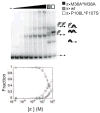
One recurring theme in plasmid duplication is the recognition of the origin of replication (ori) by specific Rep proteins that bind to DNA sequences called iterons. For plasmid R6K, this process involves a complex interplay between monomers and dimers of the Rep protein, pi, with seven tandem iterons of gamma ori. Remarkably, both pi monomers and pi dimers can bind to iterons, a new paradigm in replication control. Dimers, the predominant form in the cell, inhibit replication, while monomers facilitate open complex formation and activate the ori. Here, we investigate a mechanism by which pi monomers out-compete pi dimers for iteron binding, and in so doing activate the ori. With an in vivo plasmid incompatibility assay, we find that pi monomers bind cooperatively to two adjacent iterons. Cooperative binding is eliminated by insertion of a half-helical turn between two iterons but is diminished only slightly by insertion of a full helical turn between two iterons. These studies show also that pi bound to a consensus site promotes occupancy of an adjacent mutated site, another hallmark of cooperative interactions. pi monomer/iteron interactions were quantified using a monomer-biased pi variant in vitro with the same collection of two-iteron constructs. The cooperativity coefficients mirror the plasmid incompatibility results for each construct tested. pi dimer/iteron interactions were quantified with a dimer-biased mutant in vitro and it was found that pi dimers bind with negligible cooperativity to two tandem iterons.
Figures


Similar articles
-
Cooperative binding mode of the inhibitors of R6K replication, pi dimers.J Mol Biol. 2008 Mar 28;377(3):609-15. doi: 10.1016/j.jmb.2008.01.039. Epub 2008 Jan 26. J Mol Biol. 2008. PMID: 18295232 Free PMC article.
-
Role of pi dimers in coupling ("handcuffing") of plasmid R6K's gamma ori iterons.J Bacteriol. 2005 Jun;187(11):3779-85. doi: 10.1128/JB.187.11.3779-3785.2005. J Bacteriol. 2005. PMID: 15901701 Free PMC article.
-
Investigations of pi initiator protein-mediated interaction between replication origins alpha and gamma of the plasmid R6K.J Biol Chem. 2010 Feb 19;285(8):5695-704. doi: 10.1074/jbc.M109.067439. Epub 2009 Dec 22. J Biol Chem. 2010. PMID: 20029091 Free PMC article.
-
Plasmid R6K replication control.Plasmid. 2013 May;69(3):231-42. doi: 10.1016/j.plasmid.2013.02.003. Epub 2013 Mar 5. Plasmid. 2013. PMID: 23474464 Free PMC article. Review.
-
Regulatory implications of protein assemblies at the gamma origin of plasmid R6K - a review.Gene. 1998 Nov 26;223(1-2):195-204. doi: 10.1016/s0378-1119(98)00367-9. Gene. 1998. PMID: 9858731 Review.
Cited by
-
Toward an understanding of the DNA replication initiation in bacteria.Front Microbiol. 2024 Jan 5;14:1328842. doi: 10.3389/fmicb.2023.1328842. eCollection 2023. Front Microbiol. 2024. PMID: 38249469 Free PMC article. Review.
-
Application of the bacteriophage Mu-driven system for the integration/amplification of target genes in the chromosomes of engineered Gram-negative bacteria--mini review.Appl Microbiol Biotechnol. 2011 Aug;91(4):857-71. doi: 10.1007/s00253-011-3416-y. Epub 2011 Jun 23. Appl Microbiol Biotechnol. 2011. PMID: 21698377 Free PMC article. Review.
-
The Rep20 replication initiator from the pAG20 plasmid of Acetobacter aceti.Mol Biotechnol. 2014 Jan;56(1):1-11. doi: 10.1007/s12033-013-9680-6. Mol Biotechnol. 2014. PMID: 23839792
-
Cooperative binding mode of the inhibitors of R6K replication, pi dimers.J Mol Biol. 2008 Mar 28;377(3):609-15. doi: 10.1016/j.jmb.2008.01.039. Epub 2008 Jan 26. J Mol Biol. 2008. PMID: 18295232 Free PMC article.
-
Replisome Assembly at Bacterial Chromosomes and Iteron Plasmids.Front Mol Biosci. 2016 Aug 11;3:39. doi: 10.3389/fmolb.2016.00039. eCollection 2016. Front Mol Biosci. 2016. PMID: 27563644 Free PMC article. Review.
References
-
- Krüger R, Rakowski SA, Filutowicz M. Participating elements in the replication of iteron-containing plasmids (ICPs) In: Funnell BE, Phillips GJ, editors. Plasmid Biology. ASM Press; Washington, DC: 2004.
-
- Chen D, Feng J, Krüger R, Urh M, Inman RB, Filutowicz M. Replication of R6K γ origin in vitro: discrete start sites for DNA synthesis dependent on π and its copy-up variants. J Mol Biol. 1998;282:775–87. - PubMed
Publication types
MeSH terms
Substances
Grants and funding
LinkOut - more resources
Full Text Sources
Research Materials
Miscellaneous

