

Stereologic analysis of the lateral geniculate nucleus of the thalamus in normal and schizophrenic subjects
- PMID: 17383740
- PMCID: PMC2048985
- DOI: 10.1016/j.psychres.2006.11.003
Stereologic analysis of the lateral geniculate nucleus of the thalamus in normal and schizophrenic subjects
Abstract
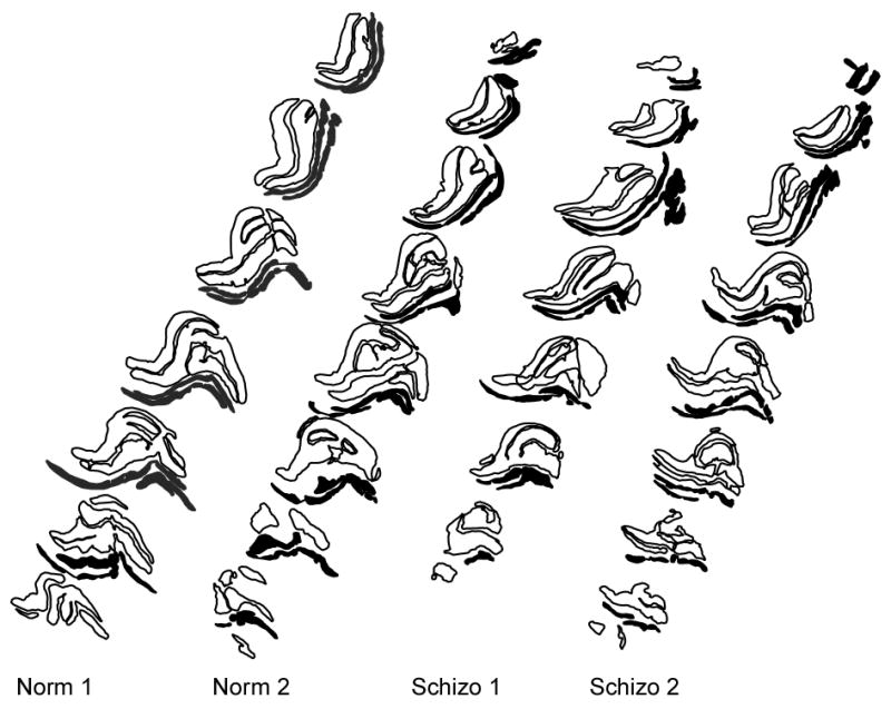
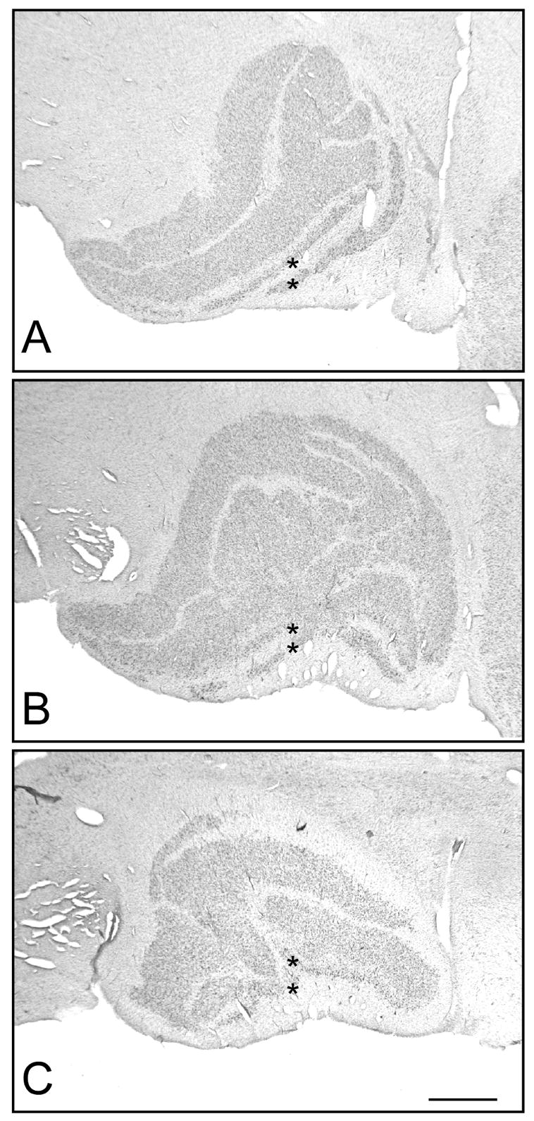
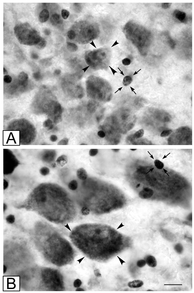
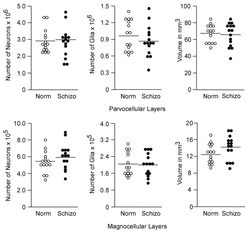
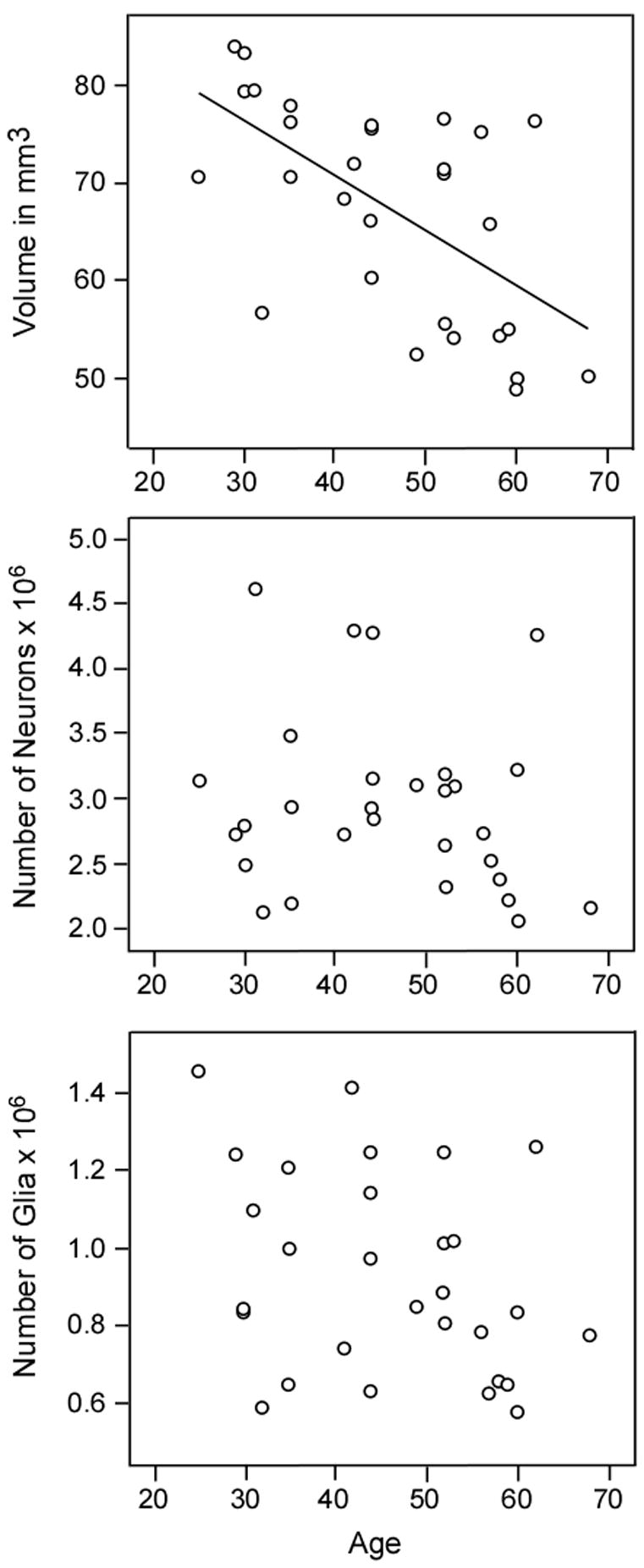
Reduction of volume and neuronal number has been found in several association nuclei of the thalamus in schizophrenic subjects. Recent evidence suggests that schizophrenic patients exhibit abnormalities in early visual processing and that many of the observed perceptual deficits are consistent with dysfunction of the magnocellular pathway, i.e. the visual relay from peripheral retinal cells to the two ventrally located magnocellular layers of the lateral geniculate nucleus (LGN). The present study was undertaken to determine whether abnormalities in cell number and volume of the LGN are associated with schizophrenia and whether the structural alterations are restricted to either the magnocellular or parvocellular subdivisions of the LGN. Series of Nissl-stained sections spanning the LGN were obtained from 15 schizophrenic and 15 normal control subjects. The optical disector/fractionator sampling method was used to estimate total neuronal number, total glial number and volume of the magnocellular and parvocellular subdivisions of the LGN. Cell number and volume of the LGN in schizophrenic subjects were not abnormal. Volume of both parvocellular and magnocellular layers of the LGN decreased with age. These findings do not support the hypothesis that early visual processing deficits in schizophrenic subjects are due to reduction of neuronal number in the LGN.
Figures
References
-
- Ahmad A, Spear PD. Effects of aging on the size, density and number of rhesus monkey lateral geniculate neurons. Journal of Comparative Neurology. 1993;3334:631–643. - PubMed
-
- Andreasen NC, Arndt S, Swayze V, 2nd, Cizaldo T, Flaum M, O’Leary D, Ehrnardt JC, Yuh WT. Thalamic abnormalities in schizophrenia visualized through magnetic resonance image averaging. Science. 1994;266:294–298. - PubMed
-
- Arciniegas D, Rojas DC, Teale P, Sheeder J, Sandberg E, Reite M. The thalamus and schizophrenia phenotype: Failure to replicate reduced volume. Biological Psychiatry. 1999;45:1329–1335. - PubMed
-
- Barch DM, Carter CS, Braver TS, Sabb FN, MacDonald A, 3rd, Noll DC, Cohen JD. Selective deficits in prefrontal cortex function in medication-naïve patients with schizophrenia. Archives of General Psychiatry. 2001;58:280–288. - PubMed
Publication types
MeSH terms
Grants and funding
LinkOut - more resources
Full Text Sources
Medical
