

doi: 10.1016/j.neuroimage.2007.02.019.
Epub 2007 Feb 23.
Detection and measurement of neurofibromatosis-1 mouse optic glioma in vivo
Affiliations
- PMID: 17383899
- PMCID: PMC2735870
- DOI: 10.1016/j.neuroimage.2007.02.019
Item in Clipboard
Detection and measurement of neurofibromatosis-1 mouse optic glioma in vivo
Neuroimage.
.
Abstract
One of the major limitations to preclinical mouse therapeutic evaluation is the inherent difficulty in imaging small tumors in vivo. We present a rapid and reliable method to detect optic glioma (OPG) in a mouse neurofibromatosis-1 model (Nf1(flox/mut)GFAP-Cre mice) in vivo using Manganese-Enhanced Magnetic Resonance Imaging (MEMRI). In a blinded study, 23 mice were chosen randomly from a cohort of Nf1(flox/mut)GFAP-Cre mice and two sets of age-matched controls. In all cases, OPG presence or absence was correctly identified. In addition, the OPG size and shape were accurately measured in vivo, facilitating the use of this model for preclinical drug studies.
Figures
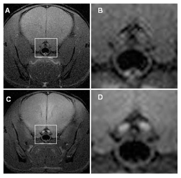
(A) Representative wild-type mouse without Mn2+ enhancement. (B) Expansion of the region marked “A” around the optic nerve (white box). (C) Same wild-type mouse with Mn2+enhancement. (D) Expansion of C around the optic nerve (white box). T1-weighted spin-echo images [repetition time (TR) = 300 ms, echo time (TE) = 14 ms, number of scans (NS) = 16] were collected in a transaxial orientation with a 1.5 × 1.5 cm2 field of view and slice thickness of 0.5 mm. Before all imaging experiments, mice were anesthetized with isoflurane/O2 [4% (v/v)], and they were maintained on isoflurane/O2 [1% (v/v)] throughout the experiments. Mouse body temperature was maintained at 37 ± 1 °C using a heating pad formed by circulating warm water.
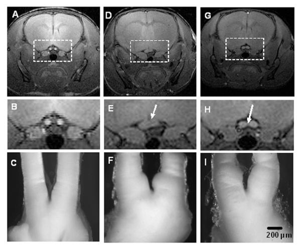
MR images and optic nerve gross pathology are shown from one representative wild-type mouse (panels A-C), and two representative Nf1flox/mutGFAP-Cre mice with optic glioma (panels D-F and G-I). The top panels show slices from T1-weighted, spin-echo MR images of the chiasmic region; the middle panels show expansions around the optic nerve region (white box); the bottom panels show corresponding the optic nerves. MEMRI was able to detect the asymmetric appearance of the optic glioma in one Nf1flox/mutGFAP-Cre mouse (panels D-F) compared to the more symmetric-appearing optic glioma in another Nf1flox/mutGFAP-Cre mouse (panels G-I).
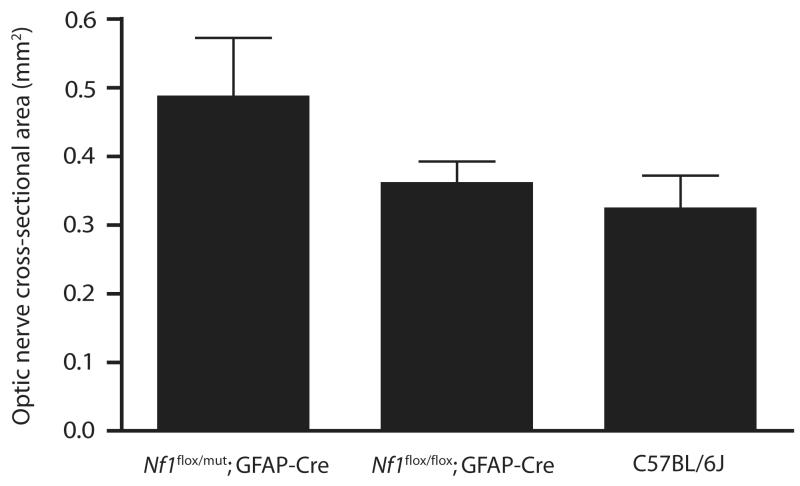
Average optic nerve cross-sectional areas in Nf1flox/mutGFAP-Cre mice (0.49 mm2; SD 0.09 mm2) are larger than those in Nf1flox/floxGFAP-Cre (0.36 mm2; SD 0.03 mm2) or C57Bl/6J mice (0.32 mm2; SD 0.05 mm2). The differences between the optic nerve areas in the Nf1flox/mutGFAP-Cre mice and the two other groups are both statistically significant; the difference between the Nf1flox/floxGFAP-Cre and C57Bl/6J mice is not statistically significant, as determined by the Mann-Whitney Rank Sum Test.
References
-
- Aoki I, Wu YJL, Silva AC, Lynch RM, Koretsky AP. In vivo detection of neuroarchitecture in the rodent brain using manganese-enhanced MRI. NeuroImage. 2004;22:1046–1059. - PubMed
-
- Bajenaru ML, Garbow JR, Perry A, Hernandez MR, Gutmann DH. Natural History of Neurofibromatosis1-Associated Optic Nerve Glioma in Mice. Ann. Neurol. 2005;57(1):119–127. - PubMed
-
- Bajenaru ML, Hernandez MR, Perry A, Y Z, Parada LF, Garbow JR, Gutmann DH. Optic Nerve Glioma in Mice Requires Astrocyte Nf1 Gene Inactivation and Nf1 Brain Heterozygosity. Cancer Res. 2003;63:8573–8577. - PubMed
-
- Listernick R, Charrow J, Greenwald M, Mets M. Natural history of optic pathway tumors in children with neurofibromatosis type 1: a longitudinal study. J. Pediatr. 1994;125:63–66. - PubMed
Publication types
MeSH terms
Substances
Grants and funding
LinkOut - more resources
Full Text Sources
Molecular Biology Databases
Research Materials
Miscellaneous
