. 2007 Jun;40(6):1544-53.
doi: 10.1016/j.bone.2007.02.012. Epub 2007 Feb 23.

Loss of myostatin (GDF8) function increases osteogenic differentiation of bone marrow-derived mesenchymal stem cells but the osteogenic effect is ablated with unloading

Affiliations

Loss of myostatin (GDF8) function increases osteogenic differentiation of bone marrow-derived mesenchymal stem cells but the osteogenic effect is ablated with unloading

M W Hamrick et al. Bone. 2007 Jun.

Abstract

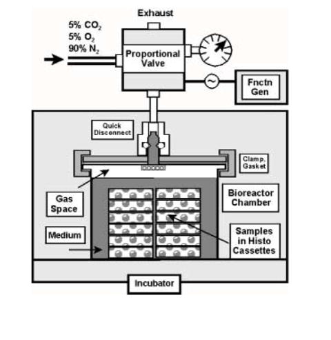
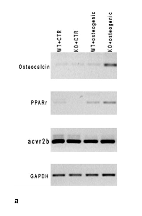
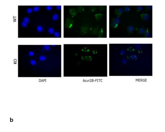
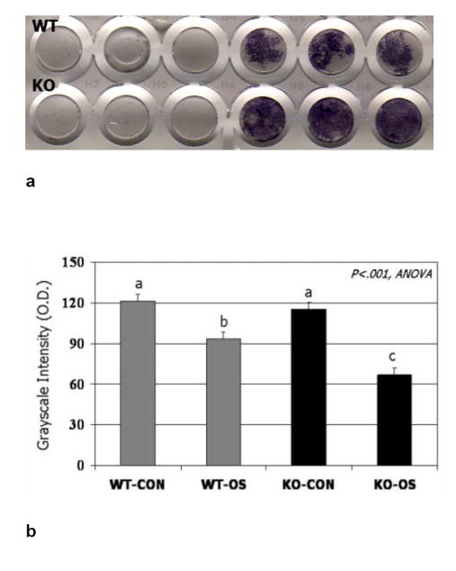
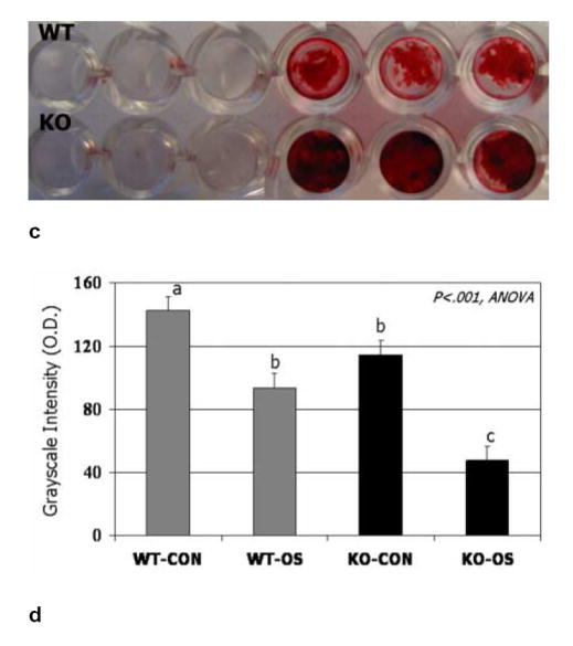
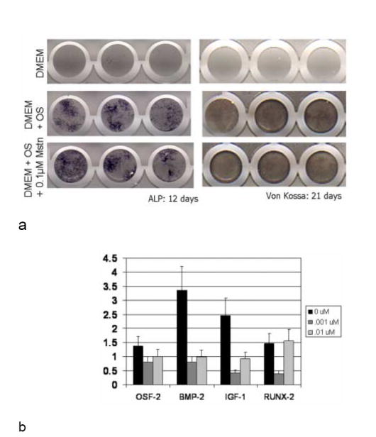
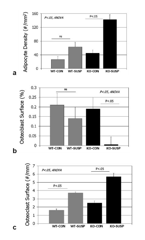
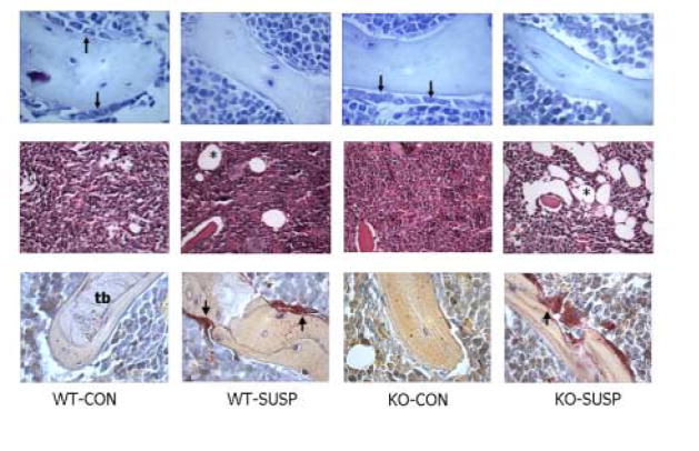
Myostatin (GDF8) is a negative regulator of skeletal muscle growth and mice lacking myostatin show a significant increase in muscle mass and bone density compared to normal mice. In order to further define the role of myostatin in regulating bone mass we sought to determine if loss of myostatin function significantly altered the potential for osteogenic differentiation in bone marrow-derived mesenchymal stem cells in vitro and in vivo. We first examined expression of the myostatin receptor, the type IIB activin receptor (AcvrIIB), in bone marrow-derived mesenchymal stem cells (BMSCs) isolated from mouse long bones. This receptor was found to be expressed at high levels in BMSCs, and we were also able to detect AcvrIIB protein in BMSCs in situ using immunofluorescence. BMSCs isolated from myostatin-deficient mice showed increased osteogenic differentiation compared to wild-type mice; however, treatment of BMSCs from myostatin-deficient mice with recombinant myostatin did not attenuate the osteogenic differentiation of these cells. Loading of BMSCs in vitro increased the expression of osteogenic factors such as BMP-2 and IGF-1, but treatment of BMSCs with recombinant myostatin was found to decrease the expression of these factors. We investigated the effects of myostatin loss-of-function on the differentiation of BMSCs in vivo using hindlimb unloading (7-day tail suspension). Unloading caused a greater increase in marrow adipocyte number, and a greater decrease in osteoblast number, in myostatin-deficient mice than in normal mice. These data suggest that the increased osteogenic differentiation of BMSCs from mice lacking myostatin is load-dependent, and that myostatin may alter the mechanosensitivity of BMSCs by suppressing the expression of osteogenic factors during mechanical stimulation. Furthermore, although myostatin deficiency increases muscle mass and bone strength, it does not prevent muscle and bone catabolism with unloading.


Figures

FIG. 1
FIG. 1
Custom bioreactor for application of hydrostatic compressive stress (pressure). Functionality of this design relies on mixed gases from a pressure cylinder to provide stress, pH balance, and support aerobic metabolism. The cyclic signal is controlled by a function generator input (upper right) to the proportional solenoid valve. Cells were plated onto glass cover slips which were positioned in histology cassettes to allow transmission of the stress to the samples.
FIG. 2
FIG. 2
a. RT-PCR results showing expression of the myostatin receptor (AcvrIIB) in BMSCs of wild-type and myostatin-deficient mice. Expression of the receptor is not altered during osteogenic culture of BMSCs. b. Immunofluorescent staining of BMSCs cultured on coverslips showing nuclear staining (DAPI, blue) and FITC-staining (green) of AcvrIIB in both wild-type and myostatin-deficient mice.
FIG. 2
FIG. 2
a. RT-PCR results showing expression of the myostatin receptor (AcvrIIB) in BMSCs of wild-type and myostatin-deficient mice. Expression of the receptor is not altered during osteogenic culture of BMSCs. b. Immunofluorescent staining of BMSCs cultured on coverslips showing nuclear staining (DAPI, blue) and FITC-staining (green) of AcvrIIB in both wild-type and myostatin-deficient mice.
FIG. 3
FIG. 3
BMSCs from wild-type and myostatin-deficient mice cultured in osteogenic medium stained after 12 days for alkaline phosphatase (ALP) activity (a) and after 21 days using alizarin red (c). Quantification shows significantly darker (low O.D. values) staining for both ALP (b) and alizarin red (d) in BMSCs from knockout mice cultured in osteogenic medium (KO-OS) compared to BMSCs from wild-type mice cultured in osteogenic medium (WT-OS). Means with different superscripts differ significantly (P<.05) in pairwise comparisons.
FIG. 3
FIG. 3
BMSCs from wild-type and myostatin-deficient mice cultured in osteogenic medium stained after 12 days for alkaline phosphatase (ALP) activity (a) and after 21 days using alizarin red (c). Quantification shows significantly darker (low O.D. values) staining for both ALP (b) and alizarin red (d) in BMSCs from knockout mice cultured in osteogenic medium (KO-OS) compared to BMSCs from wild-type mice cultured in osteogenic medium (WT-OS). Means with different superscripts differ significantly (P<.05) in pairwise comparisons.
Fig. 4
Fig. 4
a. BMSCs from myostatin-deficient mice cultured in osteogenic medium and stained for alkaline phosphatase (ALP) and Von Kossa to detect mineralized nodule formation show similar staining in the presence and absence of recombinant myostatin. Different columns of dishes represent three sample replicates. 4b. Gene expression of Mstn-/- adherent stromal cells after 3 days of cyclic hydrostatic compressive stress stimulation, consisting of 1 atmosphere at 0.5 Hz for 3 h/day. 24 h prior to testing, and between tests, cultures were incubated with 0, 10, or 100 ng/ml myostatin. Data (mean+/-SEM) are shown relative to unstressed controls which had been incubated with the same amount of myostatin. Loading increased the expression of osteogenic factors, but the addition of myostatin mitigated this trend of upregulation.
FIG. 5
FIG. 5
Histomorphometric data for wild-type cage control (wt-con), wild-type tail-suspended (wt-susp), myostatin-deficient cage control (ko-con), and myostatin-deficient tail-suspended (ko-susp). Histomorphometric variables are collected from the distal femur.
FIG. 6
FIG. 6
Marrow tissue stained with alcian blue (top row), hematoxylin and eosin (middle row) and for tartrate-resistant acid phosphatase (bottom row) in wild-type cage control (wt-con), wild-type tail-suspended (wt-susp), myostatin-deficient cage control (ko-con), and myostatin-deficient tail-suspended (ko-susp) mice. Note the large number of adipocytes (asterisks) in marrow tissue from the tail-suspended myostatin-deficient animal (middle row), and decrease in osteoblasts (arrows, top row) and increase in osteoclasts (arrows, bottom row) with tail suspension. t=trabecular bone, X200, scale bar = 0.10 mm.

References

    1. Doyle F, Brown J, Lachance C. Relation between bone mass and muscle weight. Lancet. 1970;1:391–393. - PubMed
    1. Madesen OR, Schaadt O, Bliddal H, Egsmoose C, Syvest J. Relationship between quadriceps strength and bone mineral density of the proximal tibia and distal forearm in women. J Bone Min Res. 1993;8:1439–1444. - PubMed
    1. Schönau E, Werhan E, Schniedermaier U, Mokow E, Schiessl H, Schneidhauer K, Michalk D. Influence of muscle strength on bone strength during childhood and adolescence. Horm Res. 1996;45:63–66. - PubMed
    1. Snow-Harter C, Bouxsein M, Lewis B, Charette S, Weinstein P, Marcus R. Muscle strength as a predictor of bone mineral density in young women. J Bone Miner Res. 1990;5:589–595. - PubMed
    1. Banu J, Wang L, Kalu D. Effects of increased muscle mass on bone in male mice overexpressing IGF-1 in skeletal muscles. Calcif Tissue Intl. 2003;73:196–201. - PubMed

Publication types

MeSH terms