. 2007 May;73(10):3283-90.
doi: 10.1128/AEM.02750-06. Epub 2007 Mar 23.

Assessment and interpretation of bacterial viability by using the LIVE/DEAD BacLight Kit in combination with flow cytometry

Affiliations

Assessment and interpretation of bacterial viability by using the LIVE/DEAD BacLight Kit in combination with flow cytometry

Michael Berney et al. Appl Environ Microbiol. 2007 May.

Abstract

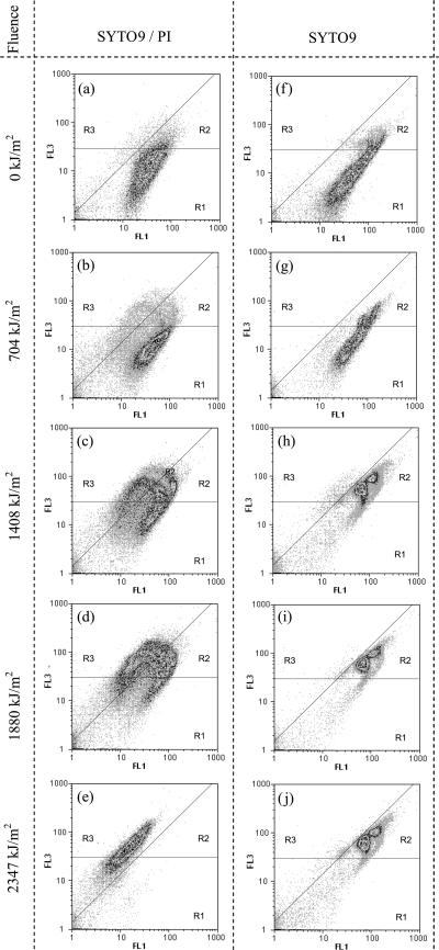
The commercially available LIVE/DEAD BacLight kit is enjoying increased popularity among researchers in various fields of microbiology. Its use in combination with flow cytometry brought up new questions about how to interpret LIVE/DEAD staining results. Intermediate states, normally difficult to detect with epifluorescence microscopy, are a common phenomenon when the assay is used in flow cytometry and still lack rationale. It is shown here that the application of propidium iodide in combination with a green fluorescent total nucleic acid stain on UVA-irradiated cells of Escherichia coli, Salmonella enterica serovar Typhimurium, Shigella flexneri, and a community of freshwater bacteria resulted in a clear and distinctive flow cytometric staining pattern. In the gram-negative bacterium E. coli as well as in the two enteric pathogens, the pattern can be related to the presence of intermediate cellular states characterized by the degree of damage afflicted specifically on the bacterial outer membrane. This hypothesis is supported by the fact that EDTA-treated nonirradiated cells exhibit the same staining properties. On the contrary, this pattern was not observed in gram-positive Enterococcus faecalis, which lacks an outer membrane. Our observations add a new aspect to the LIVE/DEAD stain, which so far was believed to be dependent only on cytoplasmic membrane permeability.


Figures

FIG. 1.
FIG. 1.
Flow cytometric analysis of E. coli K-12 MG1655 irradiated with artificial UVA light. Bacterial cells were harvested from a stationary-phase LB batch culture, washed, and diluted in mineral water (Evian). After exposure to different fluence levels (irradiation intensity × time) bacterial cell samples were stained either with a mixture of SYTO9 plus PI or with SYTO9 only and analyzed on a flow cytometer. After a fluence level of 1,408 kJ/m2 was reached, staining with SYTO9 and PI showed intermediate states (c), and both the green and red fluorescence intensity of SYTO9 increased significantly (h). After a fluence level of 2,400 kJ/m2 was reached, all cells were PI positive (e). FL1, 520 ± 10 nm; FL3, ≥630 nm.
FIG. 2.
FIG. 2.
Flow cytometric analysis of Salmonella serovar Typhimurium and S. flexneri irradiated with artificial UVA light. Bacterial cells were harvested from a stationary-phase LB batch culture, washed, and diluted in mineral water (Evian). After exposure to different fluence levels (irradiation intensity × time) bacterial cell samples were stained with a mixture of SYTO9 plus PI and analyzed on a flow cytometer (FL1, 520 ± 10 nm; FL3, ≥630 nm).
FIG. 3.
FIG. 3.
Flow cytometric analysis of E. faecalis irradiated with artificial UVA light. Bacterial cells were harvested from a stationary-phase LB batch culture, washed, and diluted in mineral water (Evian). After exposure to UVA light bacterial cell samples were stained with a mixture of SYTO9 plus PI and analyzed on a flow cytometer. R1, PI-negative cells; R2, PI-positive cells; R3, background signal. FL1, 520 ± 10 nm; FL3, ≥630 nm.
FIG. 4.
FIG. 4.
Flow cytometric analysis of E. coli K-12 MG1655 treated with 5 mM EDTA. Bacterial cells were harvested from a stationary-phase LB batch culture, washed, and diluted in mineral water (Evian). Bacterial cell samples were stained either with a mixture of SYTO9 plus PI or with SYTO9 only and analyzed on a flow cytometer. Untreated cells (a and c) and EDTA-treated cells (b and d) are compared. FL1, 520 ± 10 nm; FL3, ≥630 nm.
FIG. 5.
FIG. 5.
Comparison of a UVA-irradiated and heat-killed E. coli sample stained with SYTO9 and analyzed on the flow cytometer. Green fluorescent signals (FL1, 520 nm) are plotted against SSC signals (SSC, 488 nm). R1, single cells with single DNA content; R2, cells with double DNA content and larger average size.
FIG. 6.
FIG. 6.
Flow cytometric analysis of a community of freshwater bacteria during artificial UVA irradiation. After exposure, bacterial cell samples were stained with a mixture of SYBR green and PI and analyzed on a flow cytometer. Cells in gate R1 are PI negative. Gate R2 represents background signal. FL1, 520 ± 10 nm; FL3, ≥630 nm.

References

    1. Auty, M. A., G. E. Gardiner, S. J. McBrearty, E. O. O'Sullivan, D. M. Mulvihill, J. K. Collins, G. F. Fitzgerald, C. Stanton, and R. P. Ross. 2001. Direct in situ viability assessment of bacteria in probiotic dairy products using viability staining in conjunction with confocal scanning laser microscopy. Appl. Environ. Microbiol. 67:420-425. - PMC - PubMed
    1. Barbesti, S., S. Citterio, M. Labra, M. D. Baroni, M. G. Neri, and S. Sgorbati. 2000. Two- and three-color fluorescence flow cytometric analysis of immunoidentified viable bacteria. Cytometry 40:214-218. - PubMed
    1. Berney, M., H. U. Weilenmann, and T. Egli. 2006. Flow-cytometric study of vital cellular functions in Escherichia coli during solar disinfection (SODIS). Microbiology 152:1719-1729. - PubMed
    1. Berney, M., H. U. Weilenmann, J. Ihssen, C. Bassin, and T. Egli. 2006. Specific growth rate determines the sensitivity of Escherichia coli to thermal, UVA, and solar disinfection. Appl. Environ. Microbiol. 72:2586-2593. - PMC - PubMed
    1. Berney, M., H. U. Weilenmann, A. Simonetti, and T. Egli. 2006. Efficacy of solar disinfection of Escherichia coli, Shigella flexneri, Salmonella typhimurium and Vibrio cholerae. J. Appl. Microbiol. 101:828-836. - PubMed

Publication types

LinkOut - more resources