Review
. 2007 Apr;7(4):256-69.
doi: 10.1038/nrc2090.

Inflammation in prostate carcinogenesis

Affiliations
Review

Inflammation in prostate carcinogenesis

Angelo M De Marzo et al. Nat Rev Cancer. 2007 Apr.

Abstract

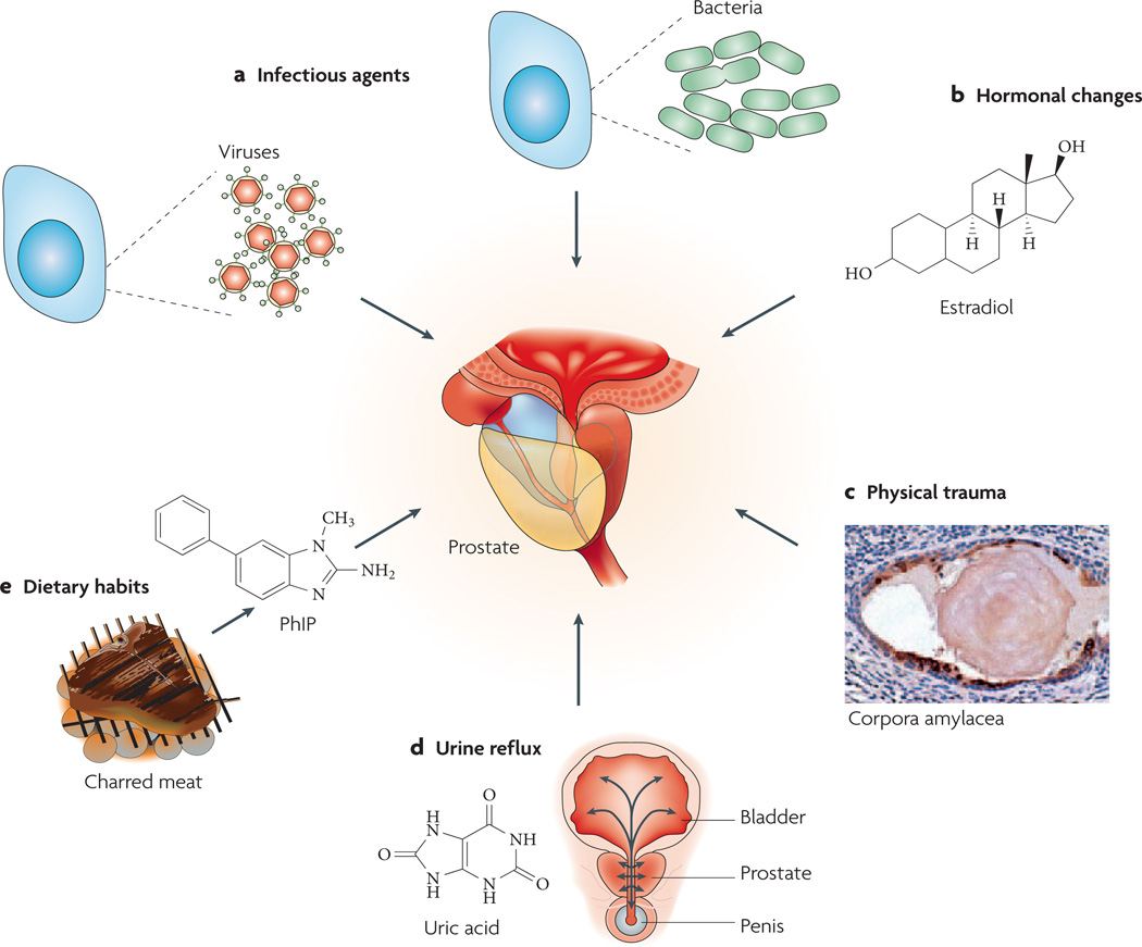
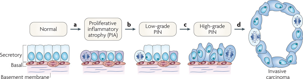
About 20% of all human cancers are caused by chronic infection or chronic inflammatory states. Recently, a new hypothesis has been proposed for prostate carcinogenesis. It proposes that exposure to environmental factors such as infectious agents and dietary carcinogens, and hormonal imbalances lead to injury of the prostate and to the development of chronic inflammation and regenerative 'risk factor' lesions, referred to as proliferative inflammatory atrophy (PIA). By developing new experimental animal models coupled with classical epidemiological studies, genetic epidemiological studies and molecular pathological approaches, we should be able to determine whether prostate cancer is driven by inflammation, and if so, to develop new strategies to prevent the disease.


Figures

Figure 1
Figure 1. Zonal predisposition to prostate disease
Most cancer lesions occur in the peripheral zone of the gland, fewer occur in the transition zone and almost none arise in the central zone. Most benign prostate hyperplasia (BPH) lesions develop in the transition zone, which might enlarge considerably beyond what is shown. The inflammation found in the transition zone is associated with BPH nodules and atrophy, and the latter is often present in and around the BPH nodules. Acute inflammation can be prominent in both the peripheral and transition zones, but is quite variable. The inflammation in the peripheral zone occurs in association with atrophy in most cases. Although carcinoma might involve the central zone, small carcinoma lesions are virtually never found here in isolation, strongly suggesting that prostatic intraepithelial neoplasia (PIN) lesions do not readily progress to carcinoma in this zone. Both small and large carcinomas in the peripheral zone are often found in association with high-grade PIN, whereas carcinoma in the transition zone tends to be of lower grade and is more often associated with atypical adenomatous hyperplasia or adenosis, and less often associated with high-grade PIN. The various patterns of prostate atrophy, some of which frequently merge directly with PIN and at times with small carcinoma lesions, are also much more prevalent in the peripheral zone, with fewer occurring in the transition zone and very few occurring in the central zone. Upper drawings are adapted from an image on Understanding Prostate Cancer website. PIN, prostatic intraepithelial neoplasia.
Figure 2
Figure 2. Possible causes of prostate inflammation
a | Infection. Chronic bacterial prostatitis is a rare recurring infection in which pathogenic bacteria are cultured from prostatic fluid. Viruses, fungi, mycobacteria and parasites can also infect the prostate and incite inflammation. The figure represents two prostate cells infected either by bacteria or viruses. b | Hormones. Hormonal alterations such as oestrogen exposure at crucial developmental junctures can result in architectural alterations in the prostate that produce an inflammatory response. c | Physical trauma. Corpora amylacea can traumatize the prostate on a microscopic level. The figure shows a corpora within a prostatic acinus in which its edges appear to be eroding the epithelium, resulting in an increase in expression of the stress enzyme cyclooxygenase 2 (PTGS2), represented by brown immunostaining. Prostate cell nuclei are visible in violet following haematoxylin staining. d | Urine reflux. Urine that travels up back towards the bladder (‘retrograde’ movement) can penetrate the ducts and acini of the prostate. Some compounds, such as crystalline uric acid, can directly activate innate inflammatory cells. Although these compounds would not be expected to traverse the prostate epithelium, if the epithelium was already damaged this would facilitate the leakage of these compounds into the stromal space where they would readily activate inflammatory cells. e | Dietary habits. Ingested carcinogens (for example 2-amino-1-methyl-6-phenylimidazo[4,5-b]pyridine (PhIP), which derives from charred meat) can reach the prostate through the bloodstream or by urine reflux and cause DNA damage and mutations, and result in an influx of inflammatory cells.
Figure 3
Figure 3. Cellular and molecular model of early prostate neoplasia progression
a | This stage is characterized by the infiltration of lymphocytes, macrophages and neutrophils (caused either by repeated infections, dietary factors and/or by the onset of autoimmunity); phagocytes release reactive oxygen and nitrogen species causing DNA damage, cell injury and cell death, which trigger the onset of epithelial cell regeneration. The morphological manifestation of the cellular injury is focal prostate atrophy, which is proposed to signify the ‘field effect’ in the prostate. The downregulation of p27, NKX3.1 and phosphatase and tensin homologue (PTEN) proteins in luminal cells stimulates cell-cycle progression. Stress-response genes are induced (such as glutathione S-transferase P1 (GSTP1), GSTA1 and cyclooxygenase 2 (PTGS2)). b | The subsequent silencing of GSTP1 through promoter methylation in subsets of cells further facilitates oxidant-mediated telomere shortening. c | Cells carrying methylated GSTP1 alleles and short telomeres have dysfunctional telomeres and are more likely to bypass the senescence checkpoints. This favours the onset of genetic instability and the consequent accumulation of genetic changes (for example loss of heterozygosity on 8p21,6q or gain of function on 8q24,17q). d | The continued proliferation of genetically unstable luminal cells and the further accumulation of genomic changes, such as gene rearrangements leading to TMPRSS2–ETS family member gene fusions, lead to progression towards invasive carcinomas. PIN, prostatic intraepithelial neoplasia.

Similar articles

Cited by

References

    1. Jemal A, et al. Cancer statistics, 2005. CA Cancer J. Clin. 2005;55:10–30. - PubMed
    1. Ames BN, Gold LS, Willett WC. The causes and prevention of cancer. Proc. Natl Acad. Sci. USA. 1995;92:5258–5265. This paper describes the main environmental causes of cancer and the molecular mechanisms by which they function.

    1. Coussens LM, Werb Z. Inflammation and cancer. Nature. 2002;420:860–867. - PMC - PubMed
    1. ACS. Cancer Facts and FIGS 2005. American Cancer Society. 2005:1–64.
    1. De Marzo AM, et al. Pathological and molecular mechanisms of prostate carcinogenesis: implications for diagnosis, detection, prevention, and treatment. J. Cell Biochem. 2004;91:459–477. - PubMed

Publication types