. 2007 Jun;148(3):564-72.
doi: 10.1111/j.1365-2249.2007.03372.x. Epub 2007 Mar 26.

In vitro treatment of monocytes with 8-methoxypsolaren and ultraviolet A light induces dendritic cells with a tolerogenic phenotype

Affiliations

In vitro treatment of monocytes with 8-methoxypsolaren and ultraviolet A light induces dendritic cells with a tolerogenic phenotype

A Legitimo et al. Clin Exp Immunol. 2007 Jun.

Abstract

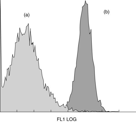
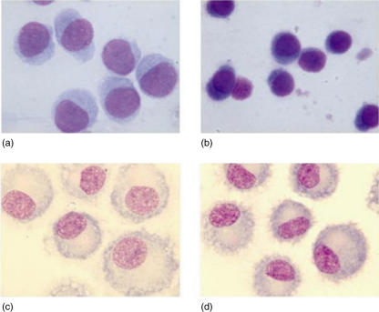
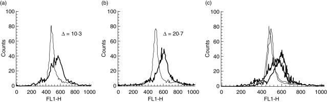
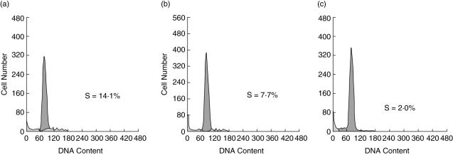
Extracorporeal photopheresis (ECP) has been considered an efficient dendritic cell (DC) therapy, used for treating both T cell malignancy, as well as T cell-mediated diseases. During the ECP procedure leucocytes are exposed to photoactivable agent 8-methoxypsolaren (8-MOP) and ultraviolet (UV) A radiation (PUVA) prior to reinfusion. Despite its clinical efficacy the mechanism of action remains elusive. As it has been reported that ECP might promote the differentiation of monocytes into immature DCs, we investigated the effects of UVA light (2 J/cm(2)) and 8-MOP (100 ng/ml) on in vitro monocyte-to-DC differentiation from normal donors. DCs were generated from human purified CD14(+) cells. Because monocytes are killed by PUVA and taking into account that only 5-10% of circulating mononuclear cells are exposed to PUVA during the ECP procedure, we developed an assay in which 10% of PUVA-treated monocytes were co-cultured with untreated monocytes. We first demonstrate that the presence of 10% apoptotic cells and monocyte activation were not enough to induce monocyte differentiation into DCs. Adding cytokines to our culture system, we obtained immature DCs characterized by significantly higher phagocytic activity and human leucocyte antigen D-related (HLA-DR) expression. These DCs preserved the capacity to be activated by lipopolysaccharide, but showed a reduced capacity to induce allogeneic T cell proliferation when first co-cultured with 10% of PUVA-treated cells. Our experimental design provides a novel insight into the real action of 8-MOP and UVA light on dendritic cell biology, suggesting an additional mechanism by which 8-MOP and UVA light exposure may influence immune responses.


Figures

Fig. 1
Fig. 1
Photoactivable agent 8-methoxypsoralen (8-MOP) and ultraviolet (UV) A (UVA) light (PUVA) treatment and plastic adherence did not induce monocyte differentiation towards dendritic cells (DCs). Photoactivable agent 8-methoxypsolaren (8-MOP) and ultraviolet (UV) A (PUVA)-exposed CD14+ cells were cultured with IL-4 and GM-CSF or without cytokines. After 6 days of culture, cells were stained with fluorescein isothiocyanate (FITC)–CD14 and analysed by flow cytometry. Plot of CD14 expression in cells generated by culturing PUVA-treated monocytes with (a) or without (b) GM-CSF/IL-4. Cells lose CD14 when cultured with IL-4 and GM-CSF (a) but they remained CD14+ in the absence of cytokines (b). Data from one representative experiment are provided.
Fig. 2
Fig. 2
The morphology of monocyte-derived dendritic cells (DCs). (a,b) Morphological profile of cells harvested during the pre-DC stage of development. Each DC preparation was examined by light microscopy (May–Grunwald–Giemsa staining). At early time-point (day 3) of culture with interleukin (IL)-4 and granulocyte–macrophage colony-stimulating factor (GM-CSF) (as described in Material and methods), cells derived from photoactivable agent 8-methoxypsolaren (8-MOP) and ultraviolet (UV) A (PUVA)-treated cultures (a) were larger in size with more cytoplasm, displaying weak basophilia, than control DCs (b). (c,d) DCs were differentiated from CD14+ cells (control Mo-DCs) cultured for 7 days with GM-CSF and IL-4. Cultures received 10 ng/ml of lipopolysaccharide (LPS) to stimulate DC maturation during the last 24 h. Cells derived from both cultures of PUVA-treated (c) and untreated monocytes (d) showed typical shapes of mature DCs such as cytoplasmic dendritic projections in both samples: the first were larger, giant-sized cells (c) in contrast to control DCs (d).
Fig. 3
Fig. 3
Photoactivable agent 8-methoxypsolaren (8-MOP) and ultraviolet (UV) A (PUVA) treatment increases the antigen uptake by dendritic cells (DCs). Cells were incubated for 30 min (at 0°C and 37°C) with fluorescein isothiocyanate (FITC)-labelled dextran, washed and analysed by flow cytometry. Representative analysis of antigen uptake by monocyte-derived (control) DCs (a) and by DCs generated from co-cultures containing PUVA-treated and untreated monocytes in the ratio 10 : 90 (b). The difference in uptake [difference in mean fluorescence intensity (ΔMFI)] between FITC–dextran uptake at 37°C (thick lines) and the control uptake at 0°C (thin lines) on day 6 is shown. Overlay of FITC–dextran uptake (c). The result is representative of eight independent experiments.
Fig. 4
Fig. 4
The mixed leucocyte reaction (MLR) stimulatory capacity of control dendritic cells (DCs) versus photoactivable agent 8-methoxypsolaren (8-MOP) and ultraviolet (UV) A radiation (PUVA)-treated DCs. Mature DCs were washed extensively and added in graded doses to allogeneic responder T cells (2 × 10⁵ cells/well) in 96-well round-bottomed microtest plates. Each group was performed in triplicate. On day 6, cells were fixed in 70% ethanol, stained with propidium iodide (10 mg/l) and processed in a flow cytometer (Epics-XL, Coulter), followed by estimating the S-phase distribution of cell cycle. Distribution of S-phase of each group: control DC stimulator cells (a), DC stimulator cells derived from co-cultures of PUVA-exposed CD14+ cells with untreated monocyte (ratio 10 : 90; see Material and methods) (b) and responder T cells only (c). The percentage of T cells in S phase is shown. Representative data are shown through one of four independent experiments with different donors.

References

    1. Ardavin C, Amigorena S, Reis e Sousa C. Dendritic cells: immunobiology and cancer immunotherapy. Immunity. 2004;20:17–23. - PubMed
    1. Usharauli D. Dendritic cells and the immunity/tolerance decision. Med Hypoth. 2005;64:112–13. - PubMed
    1. Xiao BG, Huang YM, Link H. Dendritic cell vaccine design: strategies for eliciting peripheral tolerance as therapy of autoimmune diseases. Biodrugs. 2003;17:103–11. - PubMed
    1. Schlichting CL, Schareck WD, Nickel T, Weis M. Dendritic cells as pharmacological targets for the generation of regulatory immunosuppressive effectors. New implications for allo-transplantation. Curr Med Chem. 2005;12:1921–30. - PubMed
    1. Dhodapkar MV, Steinman RM, Krasovsky J, Munz C, Bhardwaj N. Antigen-specific inhibition of effector T cell function in humans after injection of immature dendritic cells. J Exp Med. 2001;193:233–8. - PMC - PubMed

MeSH terms