. 2007 Mar 23;25(6):839-50.
doi: 10.1016/j.molcel.2007.02.003.

Requirements for Cdk7 in the assembly of Cdk1/cyclin B and activation of Cdk2 revealed by chemical genetics in human cells

Affiliations

Requirements for Cdk7 in the assembly of Cdk1/cyclin B and activation of Cdk2 revealed by chemical genetics in human cells

Stéphane Larochelle et al. Mol Cell. .

Abstract

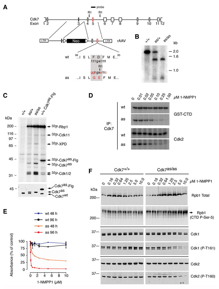
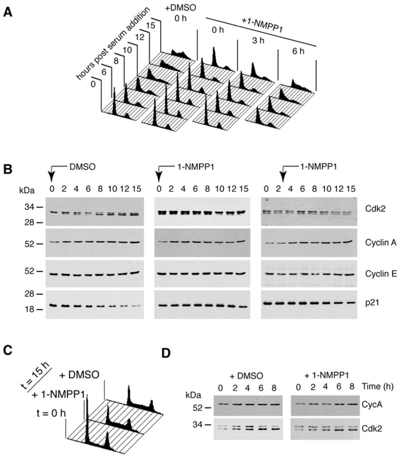
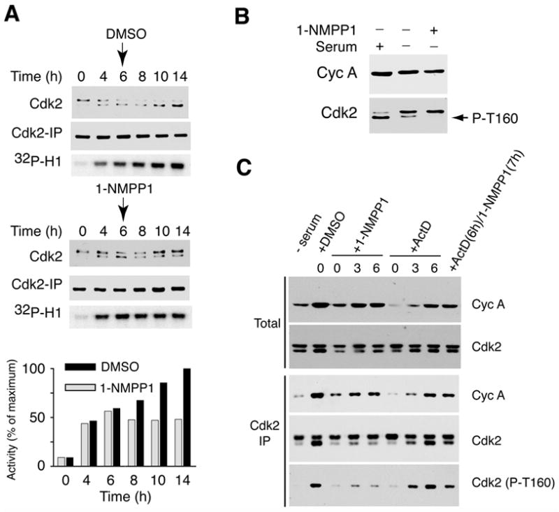
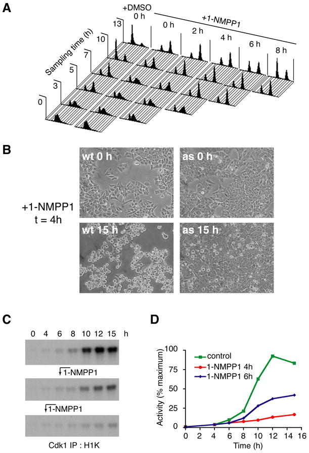
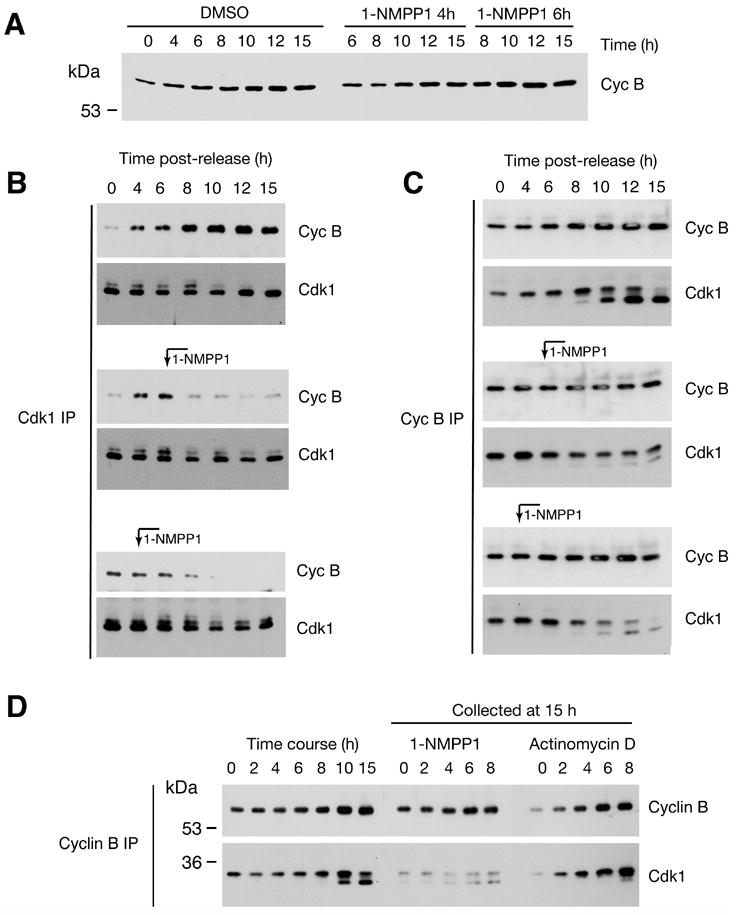
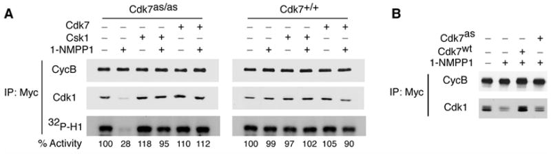
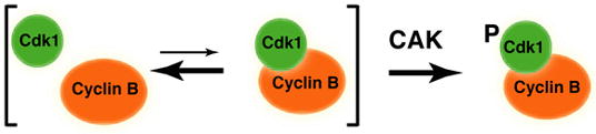
Cell division is controlled by cyclin-dependent kinases (CDKs). In metazoans, S phase onset coincides with activation of Cdk2, whereas Cdk1 triggers mitosis. Both Cdk1 and -2 require cyclin binding and T loop phosphorylation for full activity. The only known CDK-activating kinase (CAK) in metazoans is Cdk7, which is also part of the transcription machinery. To test the requirements for Cdk7 in vivo, we replaced wild-type Cdk7 with a version sensitive to bulky ATP analogs in human cancer cells. Selective inhibition of Cdk7 in G1 prevents activation (but not formation) of Cdk2/cyclin complexes and delays S phase. Inhibiting Cdk7 in G2 blocks entry to mitosis and disrupts Cdk1/cyclin B complex assembly, indicating that the two steps of Cdk1 activation-cyclin binding and T loop phosphorylation-are mutually dependent. Therefore, by combining chemical genetics and homologous gene replacement in somatic cells, we reveal different modes of CDK activation by Cdk7 at two distinct execution points in the cell cycle.


Figures

Figure 1
Figure 1. Generation of Cdk7as/as HCT116 cells
(A) The rAAV targeting vector, containing sequences from the Cdk7 locus flanking a neomycin resistance (neor) marker bounded by loxP sites. Exon 5 contained the F91G/D92E mutation and a novel EcoRI site for purposes of genotyping. The diagram indicates positions of Cdk7-derived sequences in the targeting vector and of the labeled hybridization probe, relative to EcoRI sites (RI) in the fourth and fifth introns. (B) Southern blot hybridization to EcoRI-digested genomic DNA detects single bands of ~2.0 and ~1.4 kb in the wild-type (+/+) and homozygous mutant (as/as) DNA, respectively, and an equal mixture of the two in heterozygotes (as/+). (C) Labeling with [γ-32P]N6-(benzyl)-ATP in whole-cell extracts of indicated genotypes (top). Identities of proteins labeled by both endogenous Cdk7as and recombinant, Flag-tagged Cdk7as (Cdk7as-Flg) in wild-type extract (last lane)—Rpb1 (the largest subunit of Pol II), Cdk1, -2 and -11—were inferred from similarity to the published pattern (Larochelle et al., 2006). The ~80 kDa polypeptide labeled by endogenous Cdk7as was identified by immunoprecipitation with anti-XPD antibodies. Immunoblot of kinase reaction mixtures (bottom) detects Cdk7wt and slower-migrating Cdk7as in the expected ratios; the two were expressed equally in heterozygous cells prior to excision of the neor gene (not shown), obviating the need for marker excision in homozygous Cdk7as/as cells. (D) Inhibition of Cdk7 immunoprecipitated from Cdk7as/as (AS) but not Cdk7+/+ (WT) cells by 1-NMPP1. The inhibitor was added at the indicated concentrations to GST-CTD or Cdk2 kinase assays, which contained 200 μM unlabeled ATP. (E) The dose-response of wild-type (wt) and Cdk7as/as (as) cells to 1-NMPP1 measured at 48 and 96 h of treatment by cell viability (MTT) assay. Error bars denote standard error of the mean in triplicate samples from a single representative experiment. (F) Dose-dependent effects of Cdk7 inhibition on phosphorylation of Cdk1, Cdk2 and Pol II in vivo. Asynchronous populations of Cdk7+/+ and Cdk7as/as HCT116 cells were incubated 14 h with the indicated concentrations of 1-NMPP1. Extracts were prepared and analyzed by immunoblotting with antibodies to: the amino-terminus of the Pol II subunit Rpb1 (Rpb1 Total); the Rpb1 CTD phosphorylated at the Ser5 position (CTD P-Ser-5); total Cdk1; Cdk1 phosphorylated on the T-loop (Cdk1 (P-T161)); total Cdk2 ; and Cdk2 phosphorylated on the T-loop (Cdk2 (P-T160)). Arrow at right indicates position of phosphorylated Rpb1 isoform that accumulates in Cdk7as/as cells treated with 1-NMPP1, which has a faster mobility than the slowest-migrating form (see also Supplemental Figure 1).
Figure 2
Figure 2. Inhibition of Cdk7 prevents Cdk2 activation and delays G1/S progression
(A) Cells were arrested by serum starvation, then released into serum-containing medium with DMSO or 10 μM 1-NMPP1 added at the indicated times. We monitored DNA content at indicated times by flow cytometry. (B) Levels of Cdk2, cyclin A (Cyc A), cyclin E (Cyc E) and p21 were measured in immunoblots of whole-cell extracts at indicated times after release in DMSO-treated cells, or cells treated with 1-NMPP1 0 and 3 h after release. The higher-mobility isoform of Cdk2 is phosphorylated on Thr160 of the T-loop. Its apparent absence at time 0 in the DMSO-treated cells is anomalous; the other two samples taken at time 0 are more representative (see also Figure 3). (C) Wild-type HCT116 cells were synchronized by serum starvation for 48 h, released into fresh medium containing DMSO or 1-NMPP1 as indicated and collected for flow cytometry to measure DNA content at 15 h. (D) Extracts from DMSO- or 1-NMPP1-treated, wild-type HCT116 cells, collected at indicated times after release from serum starvation, were immunoblotted with antibodies to cyclin A and Cdk2, as indicated.
Figure 3
Figure 3. Cdk7 is the Cdk2-activating kinase in vivo
(A) Addition of 1-NMPP1 6 h after serum stimulation halts Cdk2 activation. Cdk7as/as cells were released from serum starvation and harvested at indicated times for extract preparation and measurement of Cdk2 isoforms in total extract (top), and of recovered Cdk2 (middle) and histone H1 kinase activity (bottom) in anti-Cdk2 immunoprecipitates. (Cdk2 isoforms were not resolved in the immunoprecipitated samples.) DMSO or 1-NMPP1 was added as indicated at 6 h. Incorporation of 32P into histone H1 was quantified by Phosphorimager. (B) Inhibition of Cdk7 decreases Cdk2 T-loop phosphorylation in serum-starved cells. In first lane, serum-starved cells were incubated with serum for 15 h. In next two lanes, cells were treated with DMSO or 10 μM 1-NMPP1 in serum-free medium 9 h before extract preparation. (C) CAK inhibition is responsible for defective T-loop phosphorylation in 1-NMPP1-treated cells. Cells were treated with DMSO, 1-NMPP1 and/or actinomycin D (Act D), at times indicated above each lane, and harvested 15 h after release (except for first lane, time 0). Cyclin A accumulation (top) and Cdk2 levels (second from top) were measured in total lysates. Cdk2 immune complexes were isolated and probed with anti-cyclin A (third from top), anti-Cdk2 (fourth from top) or phospho-T-loop-specific (Cdk2 (T160-P), bottom) antibodies.
Figure 4
Figure 4. Cdk7 function is required for mitotic entry
(A) Execution point determination of a Cdk7-dependent function in S/G2. Cdk7as/as cells were arrested at the G1/S boundary by double-thymidine block, then released into fresh medium without or with 10 μM 1-NMPP1 added at the indicated times. To monitor cell cycle progression, we measured DNA content at indicated times by flow cytometry. Note: after release from the thymidine block, we consistently observed a minor population of cells that remained arrested with a G1 DNA content (evident, for example, in the profiles of the DMSO control culture at 3, 5 and 7 h). (B) Inhibition of Cdk7 at 4 h after release from double-thymidine block prevents mitotic entry. Wild-type (wt) and mutant (as) cells were released into nocodazole-containing medium; 1-NMPP1 was added at 4 h and cells were examined by phase-contrast microscopy at 15 h. (C) Cdk1-associated histone H1 kinase activity in cells released from G1/S (double-thymidine) block into nocodazole-containing medium. Cells were mock-treated (top), or treated with 1-NMPP1 at 6 h (middle) or 4 h (bottom) after release. (D) Incorporation of 32P into histone H1 in (C) was quantified by Phosphorimager.
Figure 5
Figure 5. Cdk7 activity is required for Cdk1/cyclin B assembly in vivo
(A) Total cyclin B levels in Cdk7as/as cells released from double-thymidine block into nocodazole-containing medium and mock-treated, or treated with 1-NMPP1 at indicated times. (B) Cdk1/cyclin B assembly was measured by immunoprecipitation with anti-Cdk1 antibodies, followed by immunoblotting with anti-cyclin B (Cyc B) and anti-Cdk1 antibodies. Where indicated, 1-NMPP1 was added at 4 or 6 h. (C) Cdk1/cyclin B assembly was measured by immunoprecipitation with anti-cyclin B (Cyc B) antibodies, followed by immunoblotting with anti-cyclin B and anti-Cdk1 antibodies. 1-NMPP1 was added at indicated time points. Cdk1 exists in two isoforms; the slower-migrating one is phosphorylated on Tyr15, and is enriched in complexes with cyclin B until cells enter mitosis. (D) Comparison of Cdk1/cyclin B binding in cells treated with 1-NMPP1 or actinomycin D. In the first seven lanes, Cdk1/cyclin B assembly was monitored at indicated times as Cdk7as/as cells progressed from a G1/S (double-thymidine) to a mitotic (nocodazole) arrest in the absence of 1-NMPP1. In the last ten lanes, 10 μM 1-NMPP1 or 10 μg/ml actinomycin D was added at indicated time after release from thymidine- into nocodazole-containing medium, and cells were harvested at 15 h.
Figure 6
Figure 6. A CAK requirement for Cdk1/cyclin B assembly recapitulated in vitro
(A) CAK is required for Cdk1/cyclin B assembly in whole-cell extracts. Mitotic extracts from Cdk7as/as or Cdk7+/+ cells were pre-incubated with or without 1-NMPP1 as indicated and supplemented with purified, recombinant Myc-tagged cyclin B. An ATP-regenerating system and, where indicated, either Cdk7/cyclin H/Mat1 complex or Csk1 was added, and extracts were incubated 90 min at room temperature. Samples were immunoprecipitated with anti-Myc antibody and immunoblotted with anti-cyclin B (CycB) or anti-Cdk1 antibodies, and tested for histone H1 kinase activity (32P-H1). Incorporation was quantified by Phosphorimager and indicated below each lane, relative to control (-1-NMPP1, -CAK) defined as 100%. (B) As in (A), except that all reactions contained Myc-tagged cyclin B added to extract from Cdk7as/as cells synchronized in G2. Where indicated, extracts were treated with 2 μM 1-NMPP1 (+) or DMSO (−) and supplemented with wild-type (Cdk7wt) or analog-sensitive (Cdk7as) CAK.
Figure 7
Figure 7. A concerted pathway of Cdk1/cyclin B assembly and activation
Cdk1 activation is a concerted reaction in vivo, with formation of a stable Cdk1/cyclin B complex occurring simultaneously with T-loop phosphorylation by Cdk7, as suggested by the interdependence of the two steps in vitro and in vivo. This pathway, if uncoupled from inhibitory mechanisms, would not generate an unphosphorylated Cdk1/cyclin B complex as an inactive intermediate.

References

    1. Abbas T, Dutta A. CDK2-Activating Kinase (CAK): More Questions than Answers. Cell Cycle. 2006;5:1123–1124. - PubMed
    1. Bishop AC, Ubersax JA, Petsch DT, Matheos DP, Gray NS, Blethrow J, Shimizu E, Tsien JZ, Schultz PG, Rose MD, et al. A chemical switch for inhibitor-sensitive alleles of any protein kinase. Nature. 2000;407:395–401. - PubMed
    1. Bornstein G, Bloom J, Sitry-Shevah D, Nakayama K, Pagano M, Hershko A. Role of the SCFSkp2 ubiquitin ligase in the degradation of p21Cip1 in S phase. J Biol Chem. 2003;278:25752–25757. - PubMed
    1. Chen J, Larochelle S, Li X, Suter B. Xpd/Ercc2 regulates CAK activity and mitotic progression. Nature. 2003;424:228–232. - PubMed
    1. Cheng A, Ross KE, Kaldis P, Solomon MJ. Dephosphorylation of cyclin-dependent kinases by type 2C protein phosphatases. Genes Dev. 1999;13:2946–2957. - PMC - PubMed

Publication types