. 2007 May;84(5):991-1000.
doi: 10.1016/j.exer.2007.01.023. Epub 2007 Feb 11.

Role of tumor invasiveness, the extracellular matrix, and chromatin sequestration in the susceptibility of uveal melanoma to herpes simplex virus type 1

Affiliations

Role of tumor invasiveness, the extracellular matrix, and chromatin sequestration in the susceptibility of uveal melanoma to herpes simplex virus type 1

Klara Valyi-Nagy et al. Exp Eye Res. 2007 May.

Abstract

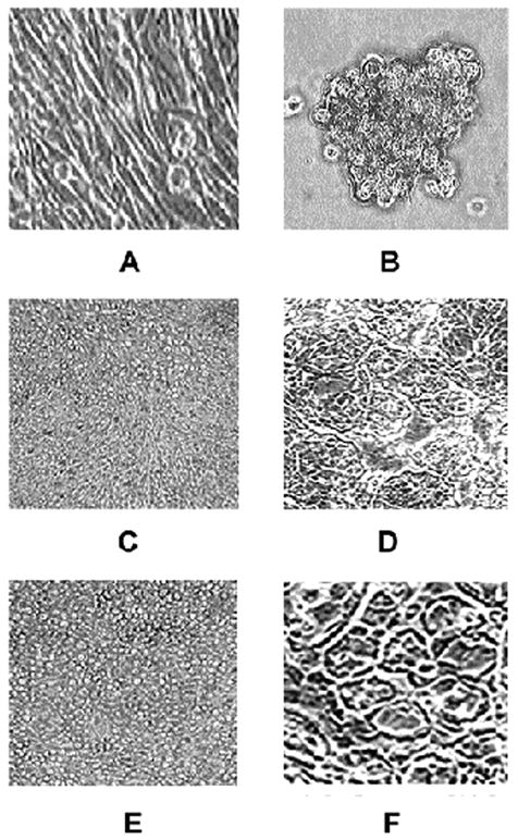
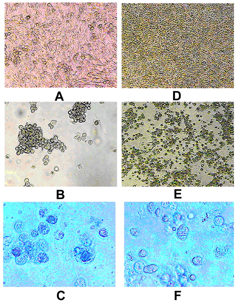
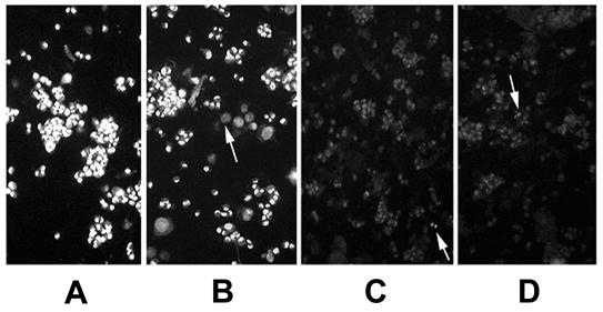
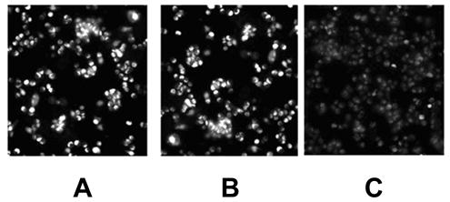
To better understand determinants of susceptibility/resistance of uveal melanomas to herpes simplex virus type 1 (HSV-1) oncolytic therapy, uveal melanoma cell lines of low (OCM1a) and of high (M619, MUM2B) invasive potential were infected with HSV-1 either in the presence or absence of a laminin-rich extracellular matrix (Matrigel). OCM1a cultures were destroyed faster by HSV-1 than M619 and MUM2B cultures. In the presence of Matrigel, all melanoma cultures demonstrated delayed destruction by HSV-1 relative to Matrigel-free cultures. As sequestration of chromatin is a characteristic feature of highly invasive uveal melanomas that is further increased by exposure to laminin, we explored whether chromatin sequestration could be reversed by HSV-1 infection. HSV-1 infection induced a global reversal of chromatin sequestration in highly invasive uveal melanoma cells. However, this viral effect was first observed only 2h following virus infection and required novel protein synthesis from input viral DNA. These findings suggest that tumor invasiveness, the spatial relationship of tumor cells to laminin and chromatin sequestration are determinants of susceptibility/resistance of melanomas to HSV-1 oncolytic therapy. Furthermore, these findings indicate for the first time that HSV-1 infection is associated with global exposure of normally highly sequestered cellular DNA in malignant cells.


Figures

Fig. 1
Fig. 1
Morphologic appearance of non-invasive and invasive uveal melanoma cells cultured in the presence or absence of laminin-rich extracellular matrix (Matrigel). OCM1a uveal melanoma cell line of low invasive potential cultured in the absence (A) or in the presence (B) of Matrigel. Highly invasive M619 uveal melanoma cells cultured in the absence (C) or in the presence (D) of Matrigel. Highly invasive MUM2B uveal melanoma cells cultured in the absence (E) or in the presence (F) of Matrigel. Note the presence of prominent vasculogenic mimicry patterns in panels D and F.
Fig. 2
Fig. 2
Morphology of mock infected and HSV-1 infected OCM1a and MUM2B melanoma cells. OCM1a and MUM2B cells were grown to approximately 70% confluency on 6-well tissue culture plates and were either exposed to 0.5 ml of sterile PBS (mock infection) or to HSV-1 diluted in PBS at a MOI=1. After incubation for 2 hours, the original inocula were removed and fresh tissue culture medium was added to each well and cultures were further incubated for times as indicated below when photographs were taken using an inverted light microscope. A. OCM1a culture 3 days after mock infection, B. OCM1a culture 3 days after HSV-1 infection, C. OCM1a culture 3 days after HSV-1 infection and following incubation of cultures with Trypan blue (0.2%) for 10 minutes, D. MUM2B culture 5 days after mock infection, E. MUM2B culture 5 days after HSV-1 infection, F. MUM2B culture 5 days after HSV-1 infection and following incubation of cultures with Trypan blue (0.2%) for 10 minutes.
Fig. 3
Fig. 3
Reversal of chromatin sequestration in MUM2B melanoma cells following HSV-1 inoculation. MUM-2 cells were grown to approximately 70% confluency on 6-well tissue culture plates. The tissue culture medium was then removed from the wells and the cells were exposed at 37° C to either 0.5 ml of sterile PBS (mock infection)(A) or to HSV-1 diluted in PBS to a final volume of 0.5 ml and a calculated MOI of 0.1 (B), 1 (C), or 10 (D) PFU/cell. After incubation for 2 hours, cells were washed twice in PBS and were then mechanically dislodged from the wells, pelleted and resuspended in 1X PBS. A drop containing 15 ul of the cell suspension was then placed on a glass slide. The drops were allowed to evaporate over a 1-hour period at room temperature. Alu I restriction enzyme (Promega) (0.5 ul in 40 ul of DMEM) was applied to the dried cells, and the preparation was placed in a humidified 37° C chamber for 2 hours when ethidium bromide was added to terminate the digestion and the preparation was photographed immediately using an inverted fluorescence microscope. Arrows in 0.1 MOI picture (B) point to the nuclei of cells that demonstrate chromatin exposure, whereas, the arrows in the 1.0 MOI (C) and the 10 MOI (D) cultures point to residual cells whose chromatin demonstrates chromatin sequestration.
Fig. 4
Fig. 4
Reversal of chromatin sequestration in MUM2B melanoma cells following HSV-2 inoculation. MUM-2 cells were grown to approximately 70% confluency on 6-well tissue culture plates. The tissue culture medium was then removed from the wells and the cells were exposed at 37° C to either 0.5 ml of sterile PBS (mock infection, B) or to HSV-2 diluted in PBS to a final volume of 0.5 ml and a MOI of 1 PFU/cell (C). After incubation for 2 hours, cells were washed twice in PBS and were then mechanically dislodged from the wells, pelleted and resuspended in 1X PBS. A drop containing 15 ul of the cell suspension was then placed on a glass slide. The drops were allowed to evaporate over a 1-hour period at room temperature. Alu I restriction enzyme (Promega) (0.5 ul in 40 ul of DMEM) was applied to the dried cells, and the preparation was placed in a humidified 37° C chamber for 2 hours when ethidium bromide was added to terminate the digestion and the preparation was photographed immediately using an inverted fluorescence microscope. Panel A shows the appearance of tumor cells without treatment (mock or HSV-2 infection) and without Alu I digestion.
Fig. 5
Fig. 5
Heat- or UV inactivated HSV-1 fails to affect chromatin exposure to Alu I digestion in highly invasive uveal melanoma cells. MUM2B cells were grown to approximately 70% confluency on 6-well tissue culture plates. The tissue culture medium was then removed from the wells and the cells were exposed at 37° C to either 0.5 ml of sterile PBS (mock infection)(A) or to HSV-1 diluted in PBS to a final volume of 0.5 ml and a MOI of 10 (B) or to UV (C) or heat-inactivated (D) HSV-1. After incubation for 3 hours, cells were washed twice in PBS and were then mechanically dislodged from the wells, pelleted and resuspended in 1X PBS. A drop containing 15 ul of the cell suspension was then placed on a glass slide. The drops were allowed to evaporate over a one-hour period at room temperature. Alu I restriction enzyme (0.5 ul in 40 ul of DMEM) was applied to the dried cells, and the preparation was placed in a humidified 37° C chamber for 2 hours when ethidium bromide was added to terminate the digestion and the preparation was photographed immediately using an inverted fluorescence microscope.

References

    1. Bedows E, Rao KM, Welsh MJ. Fate of microfilaments in Vero cells infected with measles virus and herpes simplex virus type 1. Mol Cell Biol. 1983;3:712–719. - PMC - PubMed
    1. Everett RD, Maul GG. HSV-1 IE protein Vmw110 causes redistribution of PML. EMBO J. 1994;13:5062–5069. - PMC - PubMed
    1. Everett RD, Murray J. ND10 components relocate to sites associated with herpes simplex virus type 1 nucleoprotein complexes during virus infection. J Virol. 2005;79:5078–5089. - PMC - PubMed
    1. Folberg R, Hendrix MJ, AJ Maniotis AJ. Vasculogenic mimicry and tumor angiogenesis. Am J Pathol. 2000;156:361–381. - PMC - PubMed
    1. Folberg R, Arbieva Z, Moses J, Hayee A, Sandal T, Kadkol S, Lin AK, Valyi-Nagy K, Setty S, Leach L, Chavez-Barrios P, Larsen P, Mujamdar D, Pe’er J, Maniotis AJ. Tumor cell plasticity in uveal melanoma: microenvironment directed dampening of the invasive and metastatic genotype and phenotype accompanies the generation of vasculogenic mimicry patterns. Am J Pathol. 2006;169:1376–1389. - PMC - PubMed

Publication types

MeSH terms