. 2007 Apr 23;96(8):1204-15.
doi: 10.1038/sj.bjc.6603705. Epub 2007 Mar 27.

Novel retinoic acid metabolism blocking agents have potent inhibitory activities on human breast cancer cells and tumour growth

Affiliations

Novel retinoic acid metabolism blocking agents have potent inhibitory activities on human breast cancer cells and tumour growth

J B Patel et al. Br J Cancer. .

Abstract

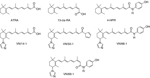
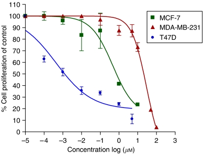
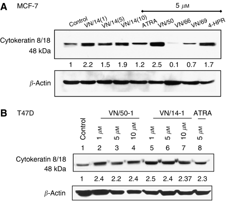
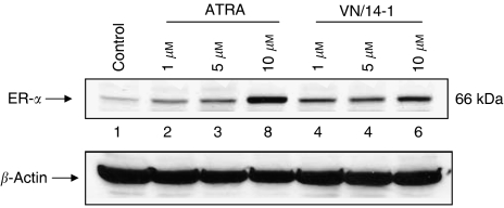
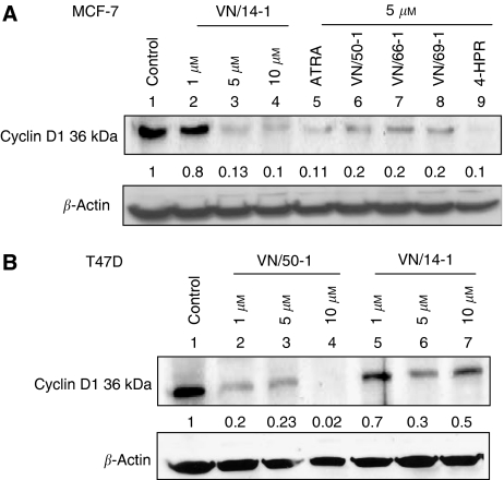
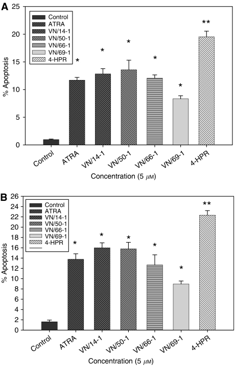
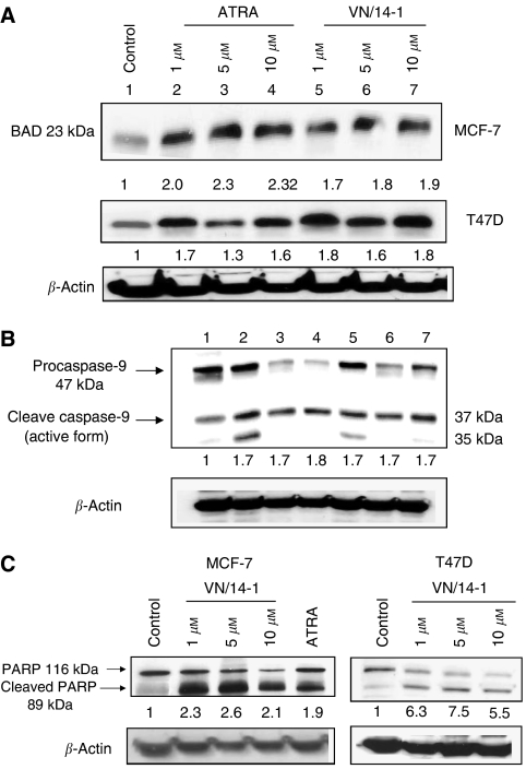
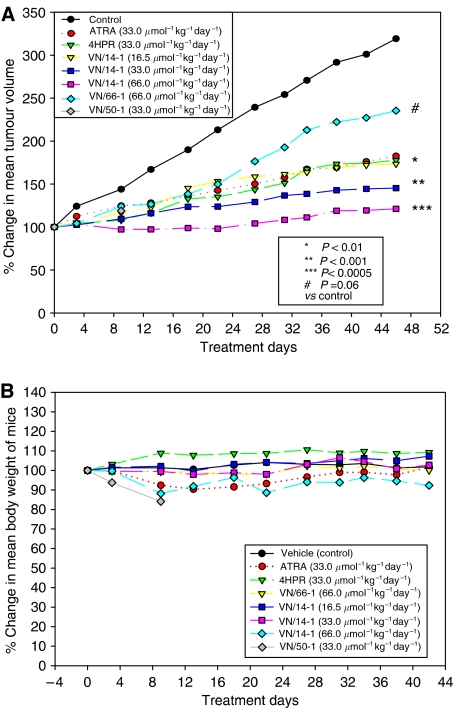
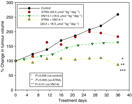
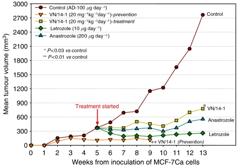
Antitumour effects of retinoids are attributed to their influence on cell proliferation, differentiation, apoptosis and angiogenesis. In our effort to develop useful agents for breast cancer therapy, we evaluated the effects of four representative retinoic acid metabolism blocking agents (RAMBAs, VN/14-1, VN/50-1, VN/66-1 and VN/69-1) on growth inhibition of oestrogen receptor positive (ER +ve, MCF-7 and T-47D) and oestrogen receptor negative (ER -ve, MDA-MB-231) human breast cancer cells. Additionally, we investigated the biological effects/molecular mechanism(s) underlying their growth inhibitory properties as well as their antitumour efficacies against MCF-7 and MCF-7Ca tumour xenografts in nude mice. We also assessed the effect of combining VN/14-1 and all-trans-retinoic acid (ATRA) on MCF-7 tumour xenografts. The ER +ve cell lines were more sensitive (IC(50) values between 3.0 and 609 nM) to the RAMBAs than the ER -ve MDA-MB-231 cell line (IC(50)=5.6-24.0 microM). Retinoic acid metabolism blocking agents induced cell differentiation as determined by increased expression of cytokeratin 8/18 and oestrogen receptor-alpha (ER-alpha). Similar to ATRA, they also induced apoptosis via activation of caspase 9. Cell cycle analysis indicated that RAMBAs arrested cells in the G1 and G2/M phases and caused significant downregulation (>80%) of cyclin D1 protein. In vivo, the growth of MCF-7 mammary tumours was dose-dependently and significantly inhibited (92.6%, P<0.0005) by VN/14-1. The combination of VN/14-1 and ATRA also inhibited MCF-7 breast tumour growth in vivo (up to 120%) as compared with single agents (P<0.025). VN/14-1 was also very effective in preventing the formation of MCF-7Ca tumours and it significantly inhibited the growth of established MCF-7Ca tumours, being as effective as the clinically used aromatase inhibitors, anastrozole and letrozole. Decrease in cyclin D1 and upregulation of cytokeratins, Bad and Bax with VN/14-1 may be responsible for the efficacy of this compound in inhibiting breast cancer cell growth in vitro and in vivo. Our results suggest that our RAMBAs, especially VN/14-1 may be useful novel therapy for breast cancer.


Figures

Figure 1
Figure 1
Structures of retinoic acids, RAMBAs and retinoid 4-HPR.
Figure 2
Figure 2
The effects of RAMBA VN/14-1 on proliferation of three human breast cancer cell line. For MCF-7 and T47D cells, 24-well plates were used and for MDA-MB-231 cells, 96-well plates were used. Cells (10 000 cells per well of a 24-well plate and 1000 cells per well of a 96-well plate) were plated and allowed to attach for 24 h. Cells were treated with VN/14-1 dissolved in 95% ethanol on day 1 and day 4 and analysed on day 7 by MTT assay using the spectrophotometer (Victor 1420 multi-label counter, Wallac (Perkin Elmer, Waltham, MA, USA)). For each concentration of the drug there were triplicate wells (24-well plate) and six wells (96-well plate) in every individual experiment. The data presented are mean±s.e.m. for 2–3 experiments. IC50 values were calculated by nonlinear regression analysis using GraphPad Prism software.
Figure 3
Figure 3
Western immunoblotting analysis of whole-cell lysates of treated MCF-7 and T47D cells for the expression of CK 8/18: (A) MCF-7 cells were treated with ATRA or RAMBAs for 6 days or 4HPR for 4 days and then cell lysates were electrophoresed using 10% SDS–PAGE and subjected to Western blotting. Lane 1: control; lanes 2–4: VN/14-1 (1, 5 and 10 μM); lane 5: ATRA (5 μM); lane 6: VN/50-1 (5 μM); lane 7: VN/66-1 (5 μM); lane 8: VN/69-1(5 μM); and lane 9: 4-HPR (5 μM). (B) T47D cells were treated with ATRA or RAMBAs (VN/50-1 and VN/14-1) for 6 days and then cell lysates were electrophoresed using 10% SDS–PAGE and subjected to Western blotting. Lane 1: control; lane 2: VN/50-1 (1 μM); lane 3: VN/50-1 (5 μM); lane 4: VN/50-1 (10 μM); lanes 5–7: VN/14-1 (1, 5 and 10 μM); and lane 8: ATRA (5 μM). Numbers below the blot show fold increase in expression of the protein as analysed by ImageQuant densitometry analysis. Membranes were stripped and probed for β-actin to verify equal protein loading. Cytokeratin 8/18 is a 48 kDa protein. Primary antibody CK 8/18 (Santa Cruz Biotechnology) 1 : 8000 in 10% milk in PBST for 1 h, and secondary antibody (anti-mouse) 1 : 2000 in 10% milk in PBST for 1 h at room temperature. The experiments were repeated thrice with similar results.
Figure 4
Figure 4
Western immunoblotting analysis of whole-cell lysates of treated MCF-7 cells for the expression of ER-α. MCF-7 cells were treated with ATRA and VN/14-1 for 6 days and then cell lysates were electrophoresed using 10% SDS–PAGE and subjected to Western blotting. Lane 1: control; lanes 2–4: ATRA (1, 5 and 10 μM); and lanes 5–7: VN/14-1 (1, 5 and 10 μM). Numbers below the blot show fold increase in expression of the protein as analysed by ImageQuant densitometry analysis. Membrane was stripped and probed for β-actin to verify equal protein loading. Primary antibody (Santa Cruz Biotechnology), 1 : 200 in 5% milk PBST for 2 h at RT, secondary antibody (anti-rabbit) 1 : 3000 in 5% milk PBST for 1 h at RT. The experiment was repeated twice with similar results.
Figure 5
Figure 5
Western blotting of whole-cell lysates of treated MCF-7 and T47D cells for the expression of Cyclin D1. (A) MCF-7 cells were treated with ATRA or RAMBAs for 6 days or 4HPR for 4 days and then cell lysates were electrophoresed using 15% SDS–PAGE and subjected to Western blotting. Lane 1: control; lanes 2–4: VN/14-1 (1, 5 and 10 μM); lane 5: ATRA (5 μM); lane 6: VN/50-1 (5 μM); lane 7: VN/66-1 (5 μM); lane 8: VN/69-1(5 μM); and lane 9: 4-HPR (5 μM). (B) T47D cells were treated with VN/50-1 and VN/14-1 for 6 days and then cell lysates were electrophoresed using 15% SDS–PAGE and subjected to Western blotting. Lane 1: control; lanes 2–4: VN/50-1 (1, 5 and 10 μM); and lanes 5–7: VN/14-1 (1, 5 and 10 μM). Numbers below the blot show fold decrease in expression of the protein as analysed by ImageQuant densitometry analysis. Membranes were stripped and probed for β-actin to verify equal protein loading. Primary antibody (Cell Signaling Technology) 1 : 2000 in 10% milk TBST overnight at 4°C and secondary antibody (anti-rabbit) 1 : 2000 in 10% milk TBST for 1 h at RT. The experiments were repeated twice with similar results.
Figure 6
Figure 6
Graphs showing apoptosis induced in (A) MCF-7 and (B) T47D cells as determined by TUNEL. (A) MCF-7 cells and (B) T47D cells (4 × 10⁴)/ well were plated in eight-well slide and treated with 5 μM of ATRA, RAMBAs and 4-HPR for 6 days (see Materials and Methods for details). TUNEL-stained apoptotic cells were counted against the DAPI stained cells to obtain percentage apoptosis. The experiments were repeated thrice with similar results. Error bars show s.e.m of three different fields of treated cells, statistically significant *P<0.01 and **P<0.001 vs control.
Figure 7
Figure 7
(A) Western immunoblotting of whole-cell lysates of treated MCF-7 and T47D cells for the expression of Bad. MCF-7 and T47D cells were treated with ATRA and VN/14-1 for 6 days and then cell lysates were electrophoresed using 15% SDS–PAGE and subjected to Western blotting. Lane 1: control; lanes 2–4: ATRA (1, 5 and 10 μM); and lanes 5–7: VN/14-1 (1 5 and 10 μM). Numbers below the blot show fold increase in expression of the protein as analysed by ImageQuant densitometry analysis. Membranes were stripped and probed for β-actin to verify equal protein loading. Primary antibody (Cell Signaling Technology) 1 : 1000 in 5% BSA TBST overnight at 4°C and secondary antibody (anti-rabbit) 1 : 2000 for 1 h at RT. This experiment was repeated twice with similar results. (B) Western blot showing activation of caspase-9 in T47D cells after treatment with ATRA or VN/14-1. T47D cells were treated with ATRA or VN/14-1 for 6 days and whole-cell lysates were electrophoresed using 10% SDS–PAGE and subjected to Western immunoblotting for caspase-9. Treatment with ATRA and VN/14-1 cleaved procaspase-9 to its active form. Lane 1: control; lanes 2–4: ATRA (1, 5 and 10 μM); and lanes 5–7: VN/14-1 (1, 5 and 10 μM). Numbers below the blot show fold increase in the active form of caspase-9 as compared with control. Membrane was stripped and probed for β-actin to verify equal amount of protein loading. The primary and secondary antibodies and their dilutions are as follows: primary antibody (Cell Signaling Technology), 1 : 1000 in 10% milk TBST overnight at 4°C and secondary antibody (anti-rabbit) 1 : 2000 in 10% milk TBST for 1 h at RT. Densitometry analysis was performed by ImageQuant software. This experiment was repeated twice with similar results. (C) Western immunoblotting of whole-cell lysates of treated MCF-7 and T47D cells for the expression of full-length and cleaved PARP. MCF-7 cells were treated with ATRA and RAMBAs for 6 days and 4-HPR for 4 days and then cell lysates were electrophoresed using 10% SDS–PAGE and subjected to Western blotting. Lane 1: control; lanes 2–4: VN/14-1 (1, 5 and 10 μM); lane 5: ATRA (5 μM); lane 6: VN/50-1 (5 μM); lane 7: VN/66-1 (5 μM); lane 8: VN/69-1(5 μM); and lane 9: 4-HPR (5 μM). T47D cells were treated with RAMBAs (VN/50-1 and VN/14-1) for 6 days and then cell lysates were electrophoresed using 10% SDS–PAGE and subjected to Western blotting. Lane 1: control; lanes 2–4: VN/50-1 (1, 5 and 10 μM); lanes 5–7: VN/14-1 (1, 5 and 10 μM). Numbers below the blot show fold increase in expression of the protein as analysed by ImageQuant densitometry analysis. Membranes were stripped and probed for β-actin to verify equal protein loading. Primary antibody (Cell Signaling Technology) 1 : 1000 in 5% milk TBST overnight at 4°C and secondary antibody (anti-rabbit) 1 : 2000 for 1 h at RT. The experiments were repeated twice with similar results.
Figure 8
Figure 8
(A) Effect of ATRA, 4HPR or RAMBAs on the growth of MCF-7 tumour xenograft in ovariectomised female athymic nude mice. Ovariectomised female athymic nude 4–6-week-old mice were used. Oestrogen pellets (1.7 mg per pellet, 90 day release obtained from Innovative Research of America) were implanted in the mice using a trochar to facilitate tumour growth. Mice were then inoculated with MCF-7 cells (2 × 10⁶ cells in Matrigel per tumour growth site) s.c. on the right and left flank. Tumours were allowed to grow for about 4–5 weeks till they were of measurable size (200–300 mm3). The mice were then grouped as control and treatment groups. Twice every week the mice were weighed and tumours were measured using a caliper. Tumour volume was calculated according to the formula 4/3πr12r2 (r1<r2). The tumour treatment study was continued for 6 weeks. (B) Effect of vehicle, ATRA 4-HPR and RAMBAs on body weight of ovariectomised female nude mice during the 6-week antitumour study. Mice were weighed twice every week during the 6-week antitumour study.
Figure 9
Figure 9
Effects of ATRA or VN/14-1 alone or in combination on the growth of MCF-7 tumour xenograft in ovariectomised female athymic nude mice. Procedure was similar to that described in Figure 8A.
Figure 10
Figure 10
Effects of VN/14-1 on the formation of MCF-7Ca tumours and effects of VN/14-1, anastrozole and letrozole on the growth of MCF-7Ca xenografts in ovariectomised female athymic nude mice. Mice were inoculated with MCF-7Ca cells as described in Materials and Methods. Beginning on the following day, androstendione (100 μg per mouse per day) was supplemented by s.c. injection for the duration of the experiment. For the tumour formation prevention group (n=5), VN/14-1 (20 mg kg−1 day−1) treatment began from the day after inoculation. For the other groups (VN/14-1, anastrazole and letrozole), treatment began after the tumour had reached approximately 300 mm3 following procedures described in Materials and Methods.

Similar articles

Cited by

References

    1. Afonja O, Juste D, Das S, Matsuhashi S, Samuels HH (2004) Induction of PDCD4 tumor suppressor gene expression by RAR agonists, antiestrogen and HER-2/neu antagonist in breast cancer cells. Evidence for a role in apoptosis. Oncogene 23: 8135–8145 - PubMed
    1. Altucci L, Gronemeyer H (2001) The promise of retinoids to fight against cancer. Nat Rev Cancer 1: 181–193 - PubMed
    1. Arapshian A, Bertran S, Kuppumbatti YS, Nakajo S, Mira-y-lopez R (2004) Epigenetic CRBP downregulation appears to be an evolutionarily conserved (human and mouse) and oncogen-specific phenomenon in breast cancer. Mol Cancer 3: 13. - PMC - PubMed
    1. Belosay A, Brodie AMH, Njar VCO (2006) Effects of novel retinoic acid metabolism blocking agent (VN/14-1) on letrozole insensitive breast cancer cells. Cancer Res 66: 11485–11493 - PubMed
    1. Bollag W, Isnardi L, Jablonska S, Klaus M, Majewski S, Pirson W, Toma S (1997) Links between pharmacological properties of retinoids and nuclear retinoid receptors. Int J Cancer 70: 470–472 - PubMed

Publication types

MeSH terms