. 2007 Apr;17(2):129-38.
doi: 10.1111/j.1750-3639.2006.00043.x.

Extensive cortical remyelination in patients with chronic multiple sclerosis

Affiliations

Extensive cortical remyelination in patients with chronic multiple sclerosis

Monika Albert et al. Brain Pathol. 2007 Apr.

Abstract

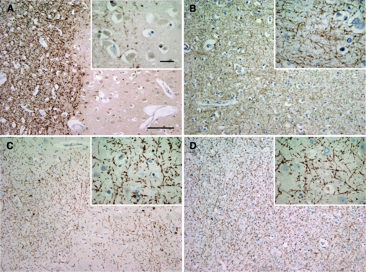
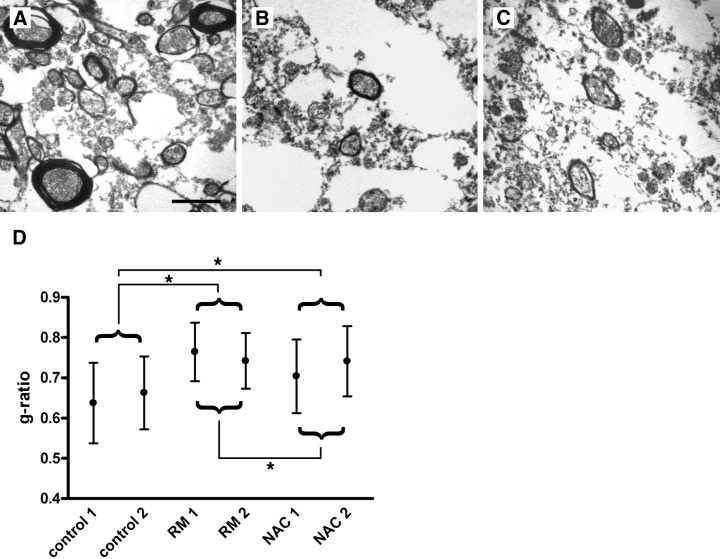
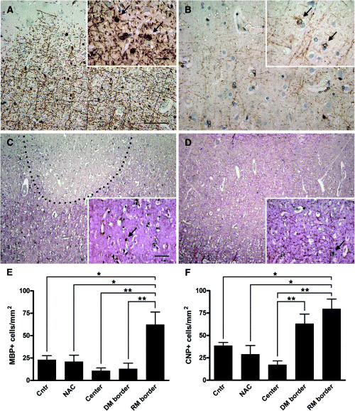
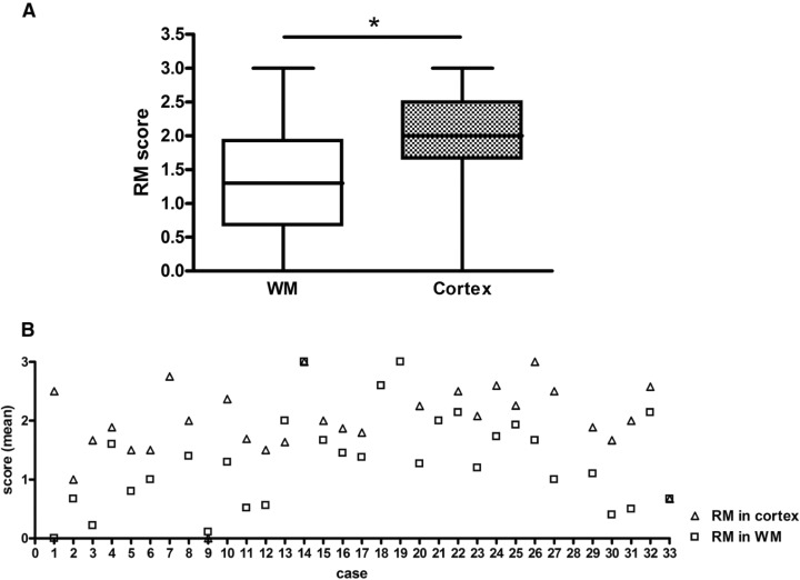
Recent studies revealed prominent cortical demyelination in patients with chronic multiple sclerosis (MS). Demyelination in white matter lesions is frequently accompanied by remyelination. This repair process, however, often remains incomplete and restricted to the lesion border. In the present study, we examined the frequency and extent of remyelination in cortical and white matter lesions in autopsy brain tissue of 33 patients with chronic MS. The majority of patients (29 of 33) harbored cortical demyelination. Remyelination of cortical lesions was identified light microscopically by the presence of thin and irregularly arranged myelin sheaths, and confirmed by electron microscopy. Extensive remyelination was found in 18%, remyelination restricted to the lesion border in 54%, and no remyelination in 28% of cortical lesions. A direct comparison of the extent of remyelination in white matter and cortical lesions of the same patients revealed that remyelination of cortical lesions was consistently more extensive. In addition, g-ratios of fibers in areas of "normal appearing cortex" yielded values consistent with remyelination. Our data confirm the high prevalence of cortical demyelination in chronic MS and imply that the propensity to remyelinate is high in cortical MS lesions.


Figures

Figure 1
Figure 1
Demyelinated and remyelinated multiple sclerosis (MS) cortex. Demyelinated cortical MS lesion. A sharp border separates well‐preserved myelin from demyelinated cortex (A). Control cortex with intact myelin (B). Cortical demyelinated lesion with patches of irregular myelin incompletely covering the lesion area (C). Normal appearing MS cortex. The myelin forms a dense mesh and is indistinguishable from non‐MS cortex (D). A–D: immunohistochemistry for myelin basic protein; blue nuclear staining: hematoxylin; scale bars: A–D: 200 µm, A–D inset: 20 µm.
Figure 2
Figure 2
Ultrastructural evidence for cortical remyelination in multiple sclerosis (MS). Representative electron micrographs of control cortex (A), a cortical lesion with light microscopical evidence for remyelination (B), and normal appearing MS cortex (C). In control cortex, myelin sheaths of regular thickness enwrap the axons (A). Thin myelin sheaths indicative of remyelination are detected in (B). Myelin sheaths of cortical fibers from areas of NAC show a thickness similar to that observed in remyelinated lesions (C). Axon to fiber (= g‐) ratios were determined in cortical lesions judged remyelinated by light microscopy [remyelination (RM); n = 2 MS patients] and normal appearing cortex (NAC; n = 2 MS patients), and compared with control cortex (n = 2). In RM and NAC cases, thinly myelinated fibers yielded higher g‐ratios as quantitative evidence for remyelination (RM vs. control: P < 0.001; NAC vs. control: P < 0.001; RM vs. NAC: P < 0.001; Kruskal–Wallis). Formalin‐fixed, paraffin‐embedded human brain tissue embedded for electron microscopy; scale bars: A–C: 2 µm.
Figure 3
Figure 3
Oligodendroglia in cortical multiple sclerosis (MS) lesions. Myelin basic protein (MBP) expressing oligodendroglia at the border of a cortical lesion (A; arrows). 2′, 3′‐cyclic nucleotide 3′‐phosphodiesterase (CNP) expressing cells in an adjacent section (B; arrows). In situ hybridization for proteolipid protein (PLP) of a cortical lesion (C) and adjacent normal appearing cortex (NAC) (D) in the same patient. Numerous PLP mRNA‐positive cells line the remyelinated border of a cortical MS lesion (C; arrow). In the NAC, only few PLP mRNA‐positive cells are detected (D; arrow). Quantitation of MBP (E) and CNP (F) positive oligodendroglial cells in demyelinated and remyelinated cortical lesions. The density of MBP‐positive oligodendrocytes is significantly higher at the remyelinated lesion border of cortical lesions compared with control cortex (P < 0.01), normal appearing cortex (P < 0.05), lesion center (P < 0.001), and border of cortical lesions without signs of remyelination (P < 0.001). Similarly, the density of CNP positive cells is significantly higher at the remyelinated lesion border of cortical lesions compared with control cortex (P < 0.01), normal appearing cortex (P < 0.01), and the lesion center (P < 0.001). Of note, many CNP‐positive cells were observed at the border of demyelinated lesions without evidence for remyelination (F). C,D: in situ hybridization for PLP mRNA (black) with immunohistochemistry for PLP protein (red); scale bars: A–D: 200 µm, A,B inset: 20 µm; C,D inset: 100 µm. Abbreviations: center = demyelinated center of lesions with and without remyelinated lesion border; cntr = control cortex; DM border = border of demyelinated lesions without evidence for remyelination; RM border = remyelinated lesion border. Statistical analysis: one‐way ANOVA with Bonferroni’s multiple comparison test.
Figure 4
Figure 4
Remyelination in white and cortical gray matter. White matter lesions (A, C, E, G) without remyelination (A), a thin rim of remyelination at the lesion border (C), substantial remyelination (E), and nearly complete remyelination (G). Cortical gray matter (B, D, F, H) with complete demyelination (B), thin remyelination at the edge of the cortical plaque (D), substantial remyelination (F) and nearly complete remyelination (H). Immunohistochemistry for myelin basic protein; blue nuclear staining: hematoxylin; scale bars: A–H: 500 µm, A–H inset: 100 µm.
Figure 5
Figure 5
Extent of cortical and white matter (WM) remyelination. The extent of cortical and WM remyelination is assessed using the semiquantitative score illustrated in Figure 4: (0) no remyelination, (1) little remyelination at the lesion border, (2) substantial remyelination, either confluent or patches of remyelination, and (3) nearly complete remyelination. 246 white and 217 gray matter lesions in the same patients were examined. Remyelination was found to be more extensive in cortical than WM lesions (A; Mann–Whitney‐U; P = 0.0013). On an individual basis, remyelination of cortical lesions was judged more extensive in 27/29 patients (open squares: WM; open triangles: cortex). Values on the y‐axis represent average scores of remyelination obtained by assessing all WM and cortical lesions of an individual patient (B). Patient 7 had no WM lesions, patients 18, 19, and 21 had no apparent cortical lesions (NAC), and patient 28 with spinal cord involvement had no lesions in the brain tissue samples available.

Similar articles

Cited by

References

    1. Allen IV (1991) Pathology of multiple sclerosis. In: Mcalpine’s Multiple Sclerosis. Matthews WB (ed.), pp. 341–378. Churchill Livingstone: Edinburgh.
    1. Baracskay KL, Duchala CS, Miller RH, Macklin WB, Trapp BD (2002) Oligodendrogenesis is differentially regulated in gray and white matter of jimpy mice. J Neurosci Res 70:645–654. - PubMed
    1. Barkhof F, Bruck W, De Groot CJ, Bergers E, Hulshof S, Geurts J, Polman CH, Van Der Valk P (2003) Remyelinated lesions in multiple sclerosis: magnetic resonance image appearance. Arch Neurol 60:1073–1081. - PubMed
    1. Barres BA, Raff MC (1993) Proliferation of oligodendrocyte precursor cells depends on electrical activity in axons. Nature 361:258–260. - PubMed
    1. Blakemore WF (1973) Remyelination of the superior cerebellar peduncle in the mouse following demyelination induced by feeding cuprizone. J Neurol Sci 20:73–83. - PubMed

Publication types