. 2007 Apr;17(2):151-64.
doi: 10.1111/j.1750-3639.2007.00058.x.

Nodule formation and desmoplasia in medulloblastomas-defining the nodular/desmoplastic variant and its biological behavior

Affiliations

Nodule formation and desmoplasia in medulloblastomas-defining the nodular/desmoplastic variant and its biological behavior

Charles S McManamy et al. Brain Pathol. 2007 Apr.

Abstract

Among the variants of medulloblastoma in the current WHO classification of nervous system tumors, the desmoplastic variant, which has been reported to constitute 5%-25% of pediatric medulloblastomas, is defined by its nodular collections of neurocytic cells bounded by desmoplastic internodular zones. We have studied the frequency, morphological features and biological behavior of medulloblastomas in two contemporaneous SIOP/UKCCSG trial cohorts of children with medulloblastomas, CNS9102 (n = 315) and CNS9204 (n = 35), focusing on tumors with nodular and desmoplastic phenotypes. In children aged 3-16 years (CNS9102), the nodular/desmoplastic medulloblastoma represented 5% of all tumors, while in infants aged <3 years (CNS9204) this variant represented 57% of medulloblastomas. Using iFISH to detect molecular cytogenetic abnormalities in medulloblastomas with a nodular architecture, we demonstrated distinct genetic profiles in desmoplastic and non-desmoplastic (classic and anaplastic) tumors; in particular, abnormalities of chromosome 17 occurred in the latter, but not the former. Significantly different outcomes were demonstrated for classic, nodular/desmoplastic and large cell/anaplastic medulloblastomas in both cohorts. In conclusion, the nodular/desmoplastic medulloblastoma appears to have clinical, genetic and biological characteristics that set it apart from other variants of this tumor.


Figures
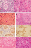
Figure 1
Figure 1
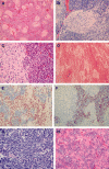
This textbook nodular/desmoplastic medulloblastoma contains nodules (A,B), in which neurocytic cells are set against a fibrillary background. Internodular regions are desmoplastic (C, reticulin preparation), and harbor cells with more nuclear pleomorphism and a higher mitotic count than intranodular cells. The border around nodules is usually sharp, but cells with an internodular phenotype can occasionally spill into nodules (D) making the border around nodules somewhat indisctinct. Differentiation along neuronal lines accompanies the neurocytic morphophenotype of intranodular cells (E, NEU‐N antibody). The growth fraction of nodules, as assessed by Ki‐67 immunolabeling, is much less than in internodular regions (F, MIB‐1 antibody). The MBEN is characterized by large nodules (G), and contrasts with the paucinodular medulloblastoma (H), in which nodules were hard to discern without the use of a reticulin preparation.
Figure 2
Figure 2
This biphasic medulloblastoma contains nodules (A–C) with the same phenotype as nodules in a nodular/desmoplastic medulloblastoma, but they lack perinodular and internodular desmoplasia (D, reticulin preparation). Intranodular tumor cells express NEU‐N and P27 (E, P27 antibody), and have a much lower Ki‐67 immunolabeling index than surrounding internodular regions (F, MIB‐1 antibody). An anaplastic phenotype characterized the internodular regions of some biphasic medulloblastomas (G). A pattern of “enlarged” rosettes was evident in a few biphasic tumors (H).
Figure 3
Figure 3
One biphasic medulloblastoma was characterized by extensive regions of neurocytic differentiation (A), in which the cell density was low and ganglion cells were occasionally found. The tumor lacked internodular desmoplasia (B, reticulin preparation). In some regions of this tumor, islands of undifferentiated cells (C), with a high mitotic count and Ki‐67 immunolabeling index (D, MIB‐1 antibody), tended to occur around small blood vessels, but were in turn surrounded by a concentric arrangement of neurocytic cells and hypocellular neurofibrillary areas (C).
Figure 4
Figure 4
This idiosyncratic biphasic medulloblastoma looked like a classic tumor in many areas, but also contained small nodules (A), without internodular desmoplasia (B, reticulin preparation), in which the condensed cells were unexpectedly characterized by a neuronal immunophenotype (C, NEU‐N antibody), and no Ki‐67 labeling (D, MIB‐1 antibody).
Figure 5
Figure 5
This and two other non‐desmoplastic medulloblastomas were characterized by foci of ganglion cell formation, and were termed ganglioneuroblastomas.
Figure 6
Figure 6
This charts the age at presentation of medulloblastoma variants. Age is plotted against the cumulative proportion of tumors in each variant’s cohort. GNB = ganglioneuroblastoma; LC/A = large cell/anaplastic; MBEN = medulloblastoma with extensive nodularity.
Figure 7
Figure 7
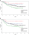
Survival curves (A—OS; B—EFS) relating to children from the CNS9102 trial for the four principal histopathological variants of medulloblastoma.

Similar articles

Cited by

References

    1. Burger PC, Grahmann FC, Bliestle A, Kleihues P (1987) Differentiation in the medulloblastoma. A histological and immunohistochemical study. Acta Neuropathol (Berl) 73:115–123. - PubMed
    1. Burger PC, Scheithauer BW (1993) Tumors of the central nervous system, 3rd edn. Atlas of Tumor Pathology, Vol. 10. Rosai J (ed.), pp. 205–217. Armed Forces Insitute of Pathology: Washington.
    1. Chatty EM, Earle KM (1971) Medulloblastoma. A report of 201 cases with emphasis on the relationship of histologic variants to survival. Cancer 28:977–983. - PubMed
    1. Collett D (1996) Modelling Survival Data in Medical Research. Chapman & Hall: London.
    1. Eberhart CG, Brat DJ, Cohen KJ, Burger PC (2000) Pediatric neuroblastic brain tumors containing abundant neuropil and true rosettes. Pediatr Dev Pathol 3:346–352. - PubMed

Publication types