. 2007 Apr;17(2):174-83.
doi: 10.1111/j.1750-3639.2007.00061.x.

The role of the octarepeat region in neuroprotective function of the cellular prion protein

Affiliations

The role of the octarepeat region in neuroprotective function of the cellular prion protein

Gerda Mitteregger et al. Brain Pathol. 2007 Apr.

Abstract

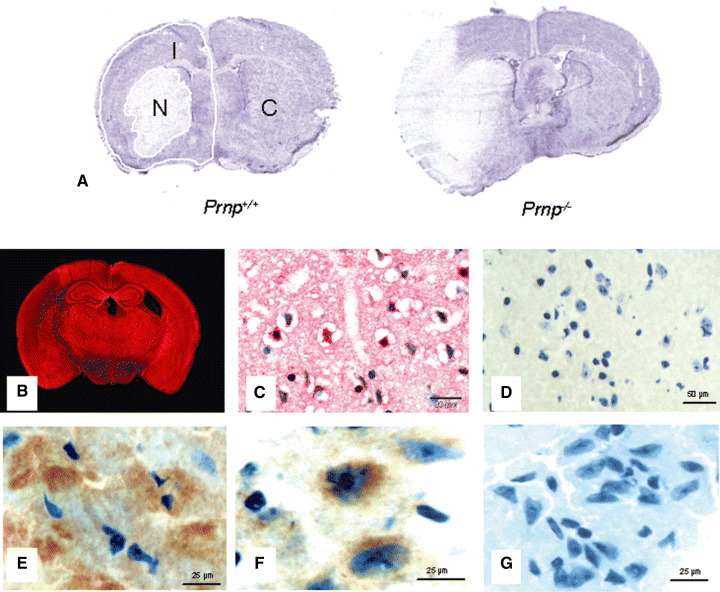
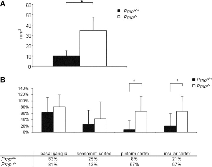
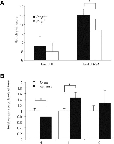
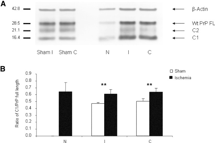
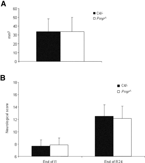
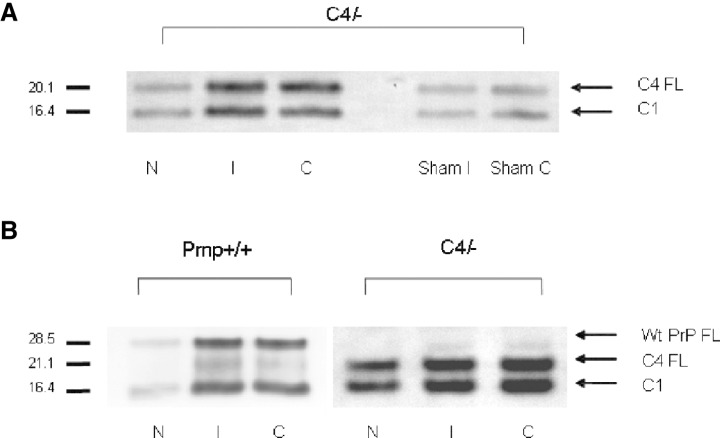
Structural alterations of the cellular prion protein (PrP(C)) seem to be the core of the pathogenesis of prion diseases. However, the physiological function of PrP(C )remains an enigma. Cell culture experiments have indicated that PrP(C) and in particular its N-terminal octarepeat region together with the phosphatidylinositol 3-kinase (PI3K)/Akt signaling pathways have a fundamental involvement in neuroprotection and oxidative stress reactions. We used wild-type mice, PrP knockout (Prnp(-/-)) animals and transgenic mice that lack the octarepeat region (C4/-) and subjected them to controlled ischemia. We identified an increased cleavage and synthesis of PrP(C) in ischemic brain areas of wild-type mice compared with sham controls. The infarct size in Prnp(-/-) animals was increased threefold when compared with wild-type mice. The infarct size in C4/- animals was identical to Prnp(-/-) mice, that is, around three times larger than in wild-type mice. We showed that the PrP in C4/- mice does not functionally rescue the Prnp(-/-) phenotype; furthermore it is unable to undergo beta cleavage, although an increased amount of C1 fragments was found in ischemic brain areas compared with sham controls. We demonstrated that the N-terminal octarepeat region has a lead function in PrP(C) physiology and neuroprotection against oxidative stress in vivo.


Figures

Figure 1
Figure 1
Histology after middle cerebral artery occlusion for 1‐h ischemia and 24‐h reperfusion time. A. Nissl‐stained sections at the level of the bregma, the maximal extent of the infarct in Prnp −/− and Prnp +/+ mice. Necrotic areas show a very faint staining. Frozen coronal sections were divided into three regions, that is, necrotic infarct area (N), ipsilateral non‐necrotic hemisphere (I) and contralateral hemisphere (C). B. Immunofluorescence for cellular prion protein (PrPC) in a Prnp +/+ mouse after cerebral ischemia using polyclonal antibody CDC1 and Fast Red as chromogen. There is strong staining for PrPC in all gray matter areas except the infarct zone on the left. C. Immunohistochemistry with polyclonal antibody CDC1. This formalin‐fixed and paraffin‐embedded section shows a number of strongly stained neurons in the area adjacent to the infarct, while other neurons and glial cells in this section appear negative. D. No reaction with the antibody directed against PrP was detected in frozen sections of the Prnp −/− littermates. E. Immunofluorescence for PrPC in Prnp +/+ mouse brains after cerebral ischemia using polyclonal antibody CDC1 and DAB as the chromogen. This frozen section shows again a number of strongly stained neurons in the area adjacent to the infarct. F. Immunofluorescence for PrPC in C4/− mouse brains after cerebral ischemia using polyclonal antibody 1A8 and DAB as the chromogen. Punctate PrP staining of intensely immunoreactive neuronal cells is seen especially in the ipsilateral area surrounding the infarct. G. No reaction with the antibody 1A8 directed against PrP was detected in the frozen sections of the Prnp −/− littermates.
Figure 2
Figure 2
A. Lesion volumes of Prnp +/+ and Prnp −/− mice after 1 h of ischemia and 24‐h reperfusion. Prnp −/− mice (n = 9) show a more than three times larger lesion volume than Prnp +/+ mice (n = 7). Data are means ± standard deviation. *P < 0.05 (Student’s t‐test). B. Neuroanatomical pattern of necrosis in Prnp +/+ and Prnp −/− mice. The graph demonstrates the extent of the infarct over various anatomical regions in the ischemic hemisphere at the level of the bregma. The affected areas in the piriform and insular cortex were significantly larger in Prnp −/− than in Prnp +/+ mice (*P < 0.05). Error bars indicate standard errors of the mean; mean values are given in the table below.
Figure 3
Figure 3
A. Neurological scores (±standard deviation) in Prnp +/+ and Prnp −/− mice subjected to transient middle cerebral artery occlusion after 1 h of ischemia (End of I1) and at the end of the 24‐h lasting reperfusion period (End of R24). No significant differences were seen after 1 h of ischemia, although there was a tendency for Prnp −/− mice to perform less well. After 24‐h reperfusion the scores of Prnp −/− mice were significantly lower than in Prnp +/+mice. The asterisk indicates significant difference (P < 0.002) as determined by t‐test. B. Quantitative reverse transcriptase polymerase chain reaction analysis of Prnp gene transcription after 1 h of transient focal cerebral ischemia and 24‐h reperfusion. The relative transcription levels of Prnp and β‐actin genes in the regions containing the necrotic infarct area (N) and the region free of infarct of the ipsilateral hemispheres (I) as well as the expression levels in the contralateral hemispheres (C) in ischemic animals are indicated by fold‐changes as compared with the corresponding regions of sham‐operated animals (n = 4 for all groups). The asterisk indicates a significant increase in Prnp transcription in the ipsilateral hemisphere I and a significant decrease in the necrotic area N compared with sham‐operated animals as determined by Student’s t‐test (P < 0.05). There is a tendency for increased Prnp transcription in the contralateral hemisphere C, which is statistically not significant.
Figure 4
Figure 4
Expression of cellular prion protein (PrPC) and generation of cleavage products in Prnp+/+ mice. A. Western blots were performed with PNGase‐digested tissue homogenates from the necrotic infarct area (N), infarct‐free brain tissue of the ipsilateral hemisphere (I) and contralateral hemisphere (C). A strong band representing full‐length PrPC (PrPC FL) was detected at about 28.5 kDa. Additional bands of lower molecular weight can be observed at about 21.1 kDa, corresponding to the C2 fragment, and at 16.4 kDa, corresponding to the C1 fragment of PrPC. As a reference the blots were reprobed with an antibody directed against β‐actin. PrPC was found decreased in necrotic areas of ischemic mouse brains, it was increased in non‐necrotic areas of the ipsilateral and contralateral hemispheres. B. Densitometric analysis of the ratio of C1/PrPC full length reveals a significant relative increase of C1 in both the necrotic infarct area (N) and the infarct‐free area of the ipsilateral hemisphere (I), and of the contralateral hemisphere (C) in ischemic mouse brains (n = 7) in comparison with areas of sham‐operated controls (n = 4). Error bars indicate standard deviations. Significant differences between ischemic and sham controls are marked by an asterisk (**P < 0.01) as determined by Student’s t‐test.
Figure 5
Figure 5
A. Lesion volumes of C4/− and Prnp −/− mice after 1 h of ischemia and 24‐h reperfusion. There are no significant differences between C4/− and Prnp −/− mice. Data are means ± standard deviation; n = 8 animals/group. B. Neurological scores (±standard deviation) in C4/− and Prnp −/− mice subjected to transient middle cerebral artery occlusion after 1 h of ischemia (End of I1) and at the end of the 24‐h lasting reperfusion period (End of R24). No significant differences in the neurological score were found at I1 and R24.
Figure 6
Figure 6
Expression of cellular prion protein (PrPC) and generation of cleavage products in C4/− and Prnp+/+ mice. The Western blots were performed with PNGase‐digested homogenates of sham‐operated C4/− mice and the three different brain regions of C4/− and Prnp +/+ mice after ischemic injury (N, I and C). A. Dominant bands of C4 full‐length (C4 FL) are detected at about 20.1 kDa in C4/− sham control mice. At about 16.4 kDa there are weaker bands that correspond to the C1 fragments in C4/− mouse brain homogenates. After middle cerebral artery occlusion (MCAO) damage in C4/− mice there is an increase in α cleavage in the three regions investigated (N, I, C). Two bands at about 20.1 and 16.4 kDa are seen corresponding to full‐length C4 protein and the C1 fragment. The signal intensities of C1 fragments in ischemic C4/− mice are remarkably stronger than those representing the C1 fragment in sham‐operated control mice. A C2 fragment is not discernible. B. A clear band of full‐length PrPC (PrPC FL) is detected at about 28.5 kDa in Prnp +/+ mice and about 20.1 kDa in C4/− mice. An additional band is seen at 21.1 kDa in the Prnp +/+ lanes corresponding to the C2 fragment. The C2 fragment of PrPC migrates approximately at the same molecular weight as the full‐length C4 protein (PrPΔ32–93). A C2 fragment is not discernible in C4/− mice. At about 16.4 kDa there are dominant bands that correspond to the C1 fragments in Prnp +/+ and C4/− mouse brain homogenates after MCAO damage.

Similar articles

Cited by

References

    1. Aronoff‐Spencer E, Burns CS, Avdievich NI, Gerfen GJ, Peisach J, Antholine WE, Ball HL, Cohen FE, Prusiner SB, Millhauser GL (2000) Identification of the Cu2+ binding sites in the N‐terminal domain of the prion protein by EPR and CD spectroscopy. Biochemistry 39:13760–13771. - PMC - PubMed
    1. Bounhar Y, Zhang Y, Goodyer CG, LeBlanc A (2001) Prion protein protects human neurons against Bax‐mediated apoptosis. J Biol Chem 276:39145–39149. - PubMed
    1. Brown DR, Qin K, Herms JW, Madlung A, Manson J, Strome R, Fraser PE, Kruck T, Von Bohlen A, Schulz‐Schaeffer W, Giese A, Westaway D, Kretzschmar H (1997) The cellular prion protein binds copper in vivo. Nature 390:684–687. - PubMed
    1. Chen SG, Teplow DB, Parchi P, Teller JK, Gambetti P, Autilio‐Gambetti L (1995) Truncated forms of the human prion protein in normal brain and in prion diseases. J Biol Chem 270:19173–19180. - PubMed
    1. Drisaldi B, Coomaraswamy J, Mastrangelo P, Strome B, Yang J, Watts JC, Chishti MA, Marvi M, Windl O, Ahrens R, Major F, Sy MS, Kretzschmar H, Fraser PE, Mount HT, Westaway D (2004) Genetic mapping of activity determinants within cellular prion proteins: N‐terminal modules in PrPC offset pro‐apoptotic activity of the Doppel helix B/B′ region. J Biol Chem 279:55443–55454. - PubMed

Publication types