

Inflammatory cell migration into the central nervous system: a few new twists on an old tale
- PMID: 17388955
- PMCID: PMC8095646
- DOI: 10.1111/j.1750-3639.2007.00067.x
Inflammatory cell migration into the central nervous system: a few new twists on an old tale
Abstract
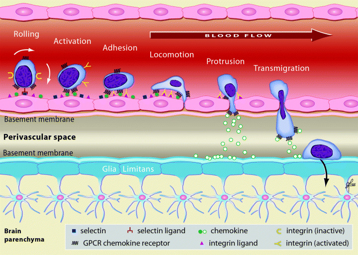
Understanding the mechanisms of leukocyte trafficking into the brain might provide insights into how to modulate pathologic immune responses or enhance host protective mechanisms in neuroinflammatory diseases such as multiple sclerosis. This review summarized our knowledge about the sites for leukocyte entry into the central nervous system, highlighting the routes from blood into the perivascular space and brain parenchyma through the blood-brain barrier. We further discussed the multistep paradigm of leukocyte-endothelial interactions at the blood-brain barrier, focusing on the adhesion molecules and chemokines involved in leukocyte transmigration. Luminal chemokines, which are immobilized on endothelial surfaces, initiate leukocyte integrin clustering and conformational change, leading to leukocyte arrest. Some leukocytes undergo post-arrest locomotion across the endothelial surface until interendothelial junctions are identified. Leukocytes then extend protrusions through the interendothelial junctions, in search of abluminal chemokines, which will serve as guidance cues for transmigration. Extravasating cells first accumulate in the perivascular space between the endothelial basement membrane and the basement membrane of the glia limitans. Matrix metalloproteases may be involved in leukocyte transverse across glia limitans into the brain parenchyma. The adhesion molecules and chemokine receptors provide attractive targets for neuroinflammatory diseases because of their important role in mediating central nervous system inflammation.
Figures
References
-
- Alon R, Grabovsky V, Feigelson S (2003) Chemokine induction of integrin adhesiveness on rolling and arrested leukocytes local signaling events or global stepwise activation? Microcirculation 10:297–311. - PubMed
-
- Alt C, Laschinger M, Engelhardt B (2002) Functional expression of the lymphoid chemokines CCL19 (ELC) and CCL 21 (SLC) at the blood‐brain barrier suggests their involvement in G‐protein‐dependent lymphocyte recruitment into the central nervous system during experimental autoimmune encephalomyelitis. Eur J Immunol 32:2133–2144. - PubMed
-
- Ancuta P, Moses A, Gabuzda D (2004) Transendothelial migration of CD16+ monocytes in response to fractalkine under constitutive and inflammatory conditions. Immunobiology 209:11–20. - PubMed
-
- Bacon KB, Harrison JK (2000) Chemokines and their receptors in neurobiology. J Neuroimmunol 104:92–97. - PubMed
Publication types
MeSH terms
Substances
LinkOut - more resources
Full Text Sources
Other Literature Sources
Medical
