. 2007 Jun;5(6):1309-17.
doi: 10.1111/j.1538-7836.2007.02527.x.

Effect of wine phenolics on cytokine-induced C-reactive protein expression

Affiliations

Effect of wine phenolics on cytokine-induced C-reactive protein expression

G Kaur et al. J Thromb Haemost. 2007 Jun.

Abstract

Background: Elevation of C-reactive protein (CRP) levels in blood was recognized as one of the cardiac disease risk factors. Consumption of wine is shown to reduce the risk from heart disease and improve longevity.

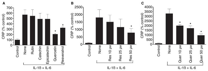
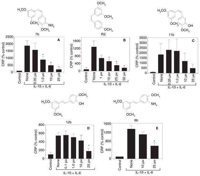
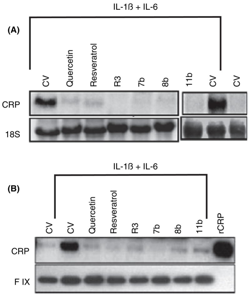
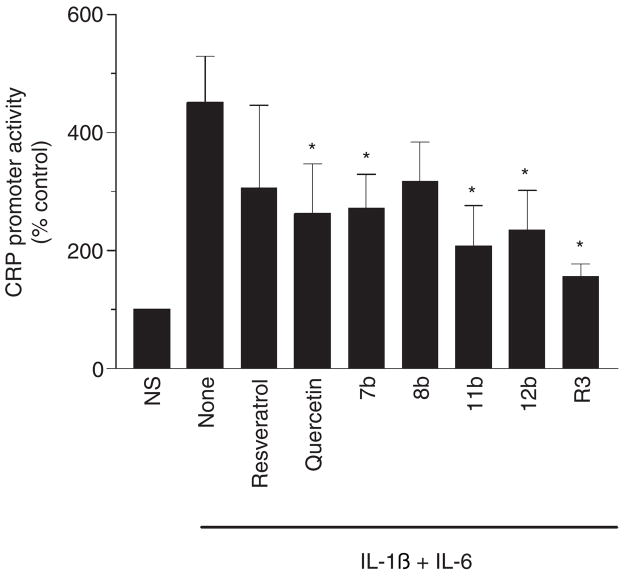
Objectives: In the present study, we evaluated the effect of various wine polyphenolic compounds and several active synthetic derivatives of resveratrol on the inflammatory cytokines (IL-1beta + IL-6)-induced CRP expression in Hep3B cells.

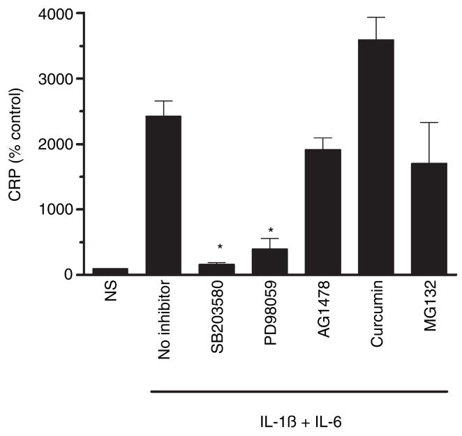
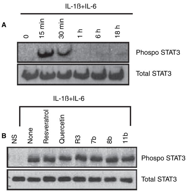
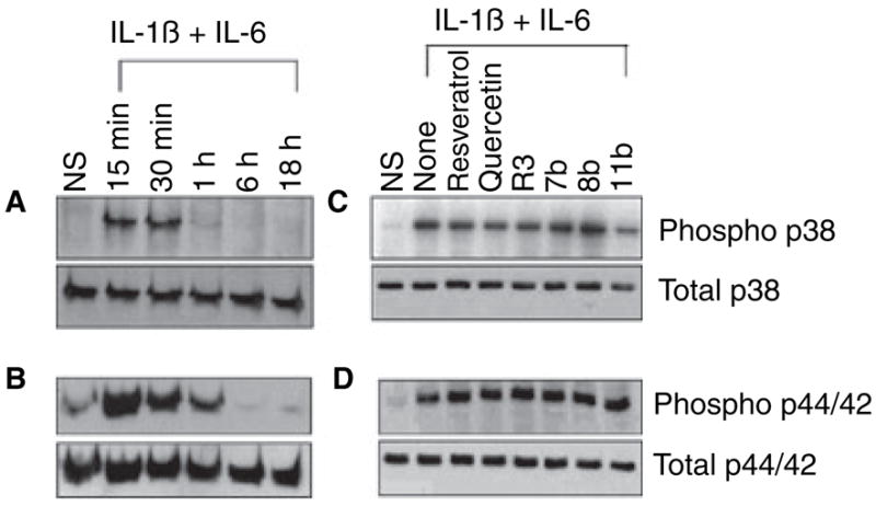
Results: Among the wine phenolics tested, quercetin and resveratrol, in a dose-dependent manner, suppressed cytokine-induced CRP expression. Two of the synthetic derivatives of resveratrol, R3 and 7b, elicited a fiftyfold higher suppressive effect compared with resveratrol. The inhibitory effects of resveratrol and its derivatives on CRP expression were at the level of mRNA production. Investigation of signaling pathways showed that the cytokines induced the phosphorylation of p38 and p44/42 MAP kinases. Inhibitors of p38 and p44/42 mitogen-activated protein kinase (MAPK) activation inhibited CRP expression, implicating the involvement of both pathways in cytokine-induced CRP expression. These data revealed a previously unrecognized role of the p44/42 MAPK signaling pathway in CRP expression. Wine polyphenolics or the synthetic compounds of resveratrol did not affect cytokine-activated phosphorylation of these MAPKs.

Conclusions: Wine phenolics inhibit CRP expression; however, to do so, they do not utilize the MAPK pathways.


Conflict of interest statement

Disclosure of Conflict of Interests

The authors state that they have no conflict of interest.

Figures

Fig. 1
Fig. 1
Effect of some major wine compounds on cytokine (IL-1β + IL-6)-induced C-reactive protein (CRP) expression. Hep3B cells were pretreated for 1 h with various wine polyphenolics (50 μM) (panel A), varying doses of resveratrol (panel B) or quercetin (panel C), and then stimulated for 24 h with a combination of IL-1β (10 ng mL−1) and IL-6 (10 ng mL−1). At the end of 24 h, the conditioned media were collected, concentrated and CRP levels were measured in ELISA. *Denotes that the CRP level in cells treated with the experimental compound was significantly lower compared with CRP levels in stimulated cells that were exposed to a control vehicle.
Fig. 2
Fig. 2
Effect of various chemically synthesized resveratrol derivatives on C-reactive protein (CRP) expression. Hep3B cells were pretreated for 1 h with varying concentrations of resveratrol derivatives and then stimulated for 24 h with a combination of IL-1β and IL-6. At the end of 24 h, the conditioned media were collected, concentrated and CRP levels were measured in ELISA. *Denotes that the CRP level in cells treated with the experimental compound was significantly lower compared with CRP levels in stimulated cells that were exposed to a control vehicle.
Fig. 3
Fig. 3
Quercetin, resveratrol and its derivatives suppress C-reactive protein (CRP) mRNA and protein synthesis. Hep3B cells were subjected to 1 h pretreatment with a control vehicle, resveratrol (50 μM), quercetin (50 μM), R3 (1 μM), 7b (1 μM), 8b (25 μM) or 11b (25 μM) in the presence of dexamethasone (1 μM). Then, the cells were stimulated with IL-1β (10 ng mL−1) and IL-6 (20 ng mL−1) for 24 h. The conditioned media were removed, concentrated and subjected to immunoblot analysis with anti-CRP antibodies (top panel B) and antifactor IX to serve as a loading control (bottom panel B). Ten ng of recombinant CRP (rCRP) protein was loaded in the last lane. The cells were extracted with Trizol reagent to obtain total RNA, and 30 μg total RNA was subjected to Northern blot analysis and probed with 32P-labeled CRP cDNA (top panel A). The 18S RNA band of the same gel was shown for loading control (bottom panel A). CV denotes control vehicle.
Fig. 4
Fig. 4
Wine phenolics inhibit transcriptional activation of C-reactive protein (CRP) promoter. Hep3B cells were transiently transfected with − 157/+9 CRP promoter region linked to luciferase reporter gene. After 6 h, the transfected cells were treated with various phenolic compounds for 1 h, and then stimulated with IL-1β and IL-6 for 24 h. The cells were extracted with reporter lysis buffer and the cell extracts were used for measurement of luciferase activity. The concentration of wine phenolics used in this study was same as in Fig. 3. NS denotes non-stimulated cells. *Denotes that the CRP promoter activity is significantly lower compared with stimulated cells that were exposed to a control vehicle.
Fig. 5
Fig. 5
Effect of various signaling pathway inhibitors on cytokine-induced C-reactive protein (CRP) expression. Hep3B cells were pretreated with a control vehicle; p38 MAPK inhibitor, SB203580 (10 μM); p44/42 MAPK inhibitor, PD98059 (10 μM); EGFR inhibitor, AG1478 (10 μM); NF-κB inhibitors, curcumin (30 μM) and MG132 (1 μM); for 1 h and then stimulated with IL-1β and IL-6 for 24 h. The conditioned media was used to measure CRP levels. NS denotes non-stimulated cells. *Denotes that the CRP levels were significantly lower compared with stimulated cells not exposed to inhibitors.
Fig. 6
Fig. 6
Wine phenolics do not suppress cytokine-induced STAT3 phosphorylation. Panel A: Hep3B cells were treated with IL-1β and IL-6 for varying time periods, and cell extracts harvested at the end of the treatment period were subjected to immunoblot analysis to evaluate STAT3 phosphorylation. Panel B: Hep3B cells were pretreated with quercetin, resveratrol or resveratrol derivatives for 1 h, and then treated with IL-1β and IL-6 for 15 min to activate STAT3. STAT3 phosphorylation was evaluated as in panel A. The concentration of wine phenolics used in this study was the same as in Fig. 3. NS denotes non-stimulated cells.
Fig. 7
Fig. 7
Effect of resveratrol and its derivatives on cytokine-induced activation of p38 and p44/42 MAPK. Hep3B cells were treated with IL-1β and IL-6 for varying time periods, and cell extracts harvested at the end of the treatment period were subjected to immunoblot analysis to evaluate p38 (Panel A) or p44/42 MAPK (panel B) activation. Panels C and D: Hep3B cells were pretreated with quercetin, resveratrol or resveratrol derivatives for 1 h, and then treated with IL-1β and IL-6 for 15 min, and the cell extracts were analyzed for p38 (panel C) and p44/42 (panel D) MAPK activation. The concentration of wine phenolics used in this study was the same as in Fig. 3.

Similar articles

Cited by

References

    1. Black S, Kushner I, Samols D. C-reactive Protein. J Biol Chem. 2004;279:48487–90. - PubMed
    1. Liuzzo G, Biasucci LM, Gallimore JR, Grillo RL, Rebuzzi AG, Pepys MB, Maseri A. The prognostic value of C-reactive protein and serum amyloid a protein in severe unstable angina. N Engl J Med. 1994;331:417–24. - PubMed
    1. Danesh J, Collins R, Appleby P, Peto R. Association of fibrinogen, C-reactive protein, albumin, or leukocyte count with coronary heart disease: meta-analyses of prospective studies. JAMA. 1998;279:1477–82. - PubMed
    1. Jialal I, Devaraj S, Venugopal SK. C-reactive protein: risk marker or mediator in atherothrombosis? Hypertension. 2004;44:6–11. - PubMed
    1. Verma S, Szmitko PE, Yeh ET. C-reactive protein: structure affects function. Circulation. 2004;109:1914–7. - PubMed

Publication types