

Novel markers for differentiation of lobular and ductal invasive breast carcinomas by laser microdissection and microarray analysis
- PMID: 17389037
- PMCID: PMC1852112
- DOI: 10.1186/1471-2407-7-55
Novel markers for differentiation of lobular and ductal invasive breast carcinomas by laser microdissection and microarray analysis
Abstract
Background: Invasive ductal and lobular carcinomas (IDC and ILC) are the most common histological types of breast cancer. Clinical follow-up data and metastatic patterns suggest that the development and progression of these tumors are different. The aim of our study was to identify gene expression profiles of IDC and ILC in relation to normal breast epithelial cells.
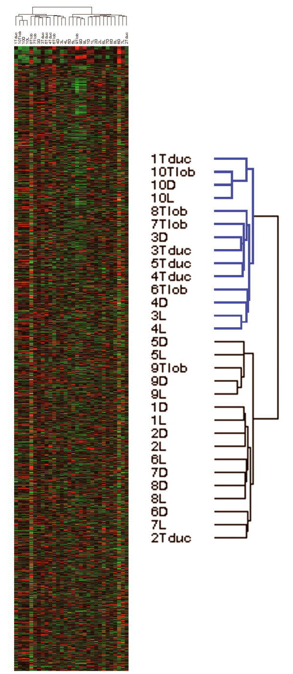
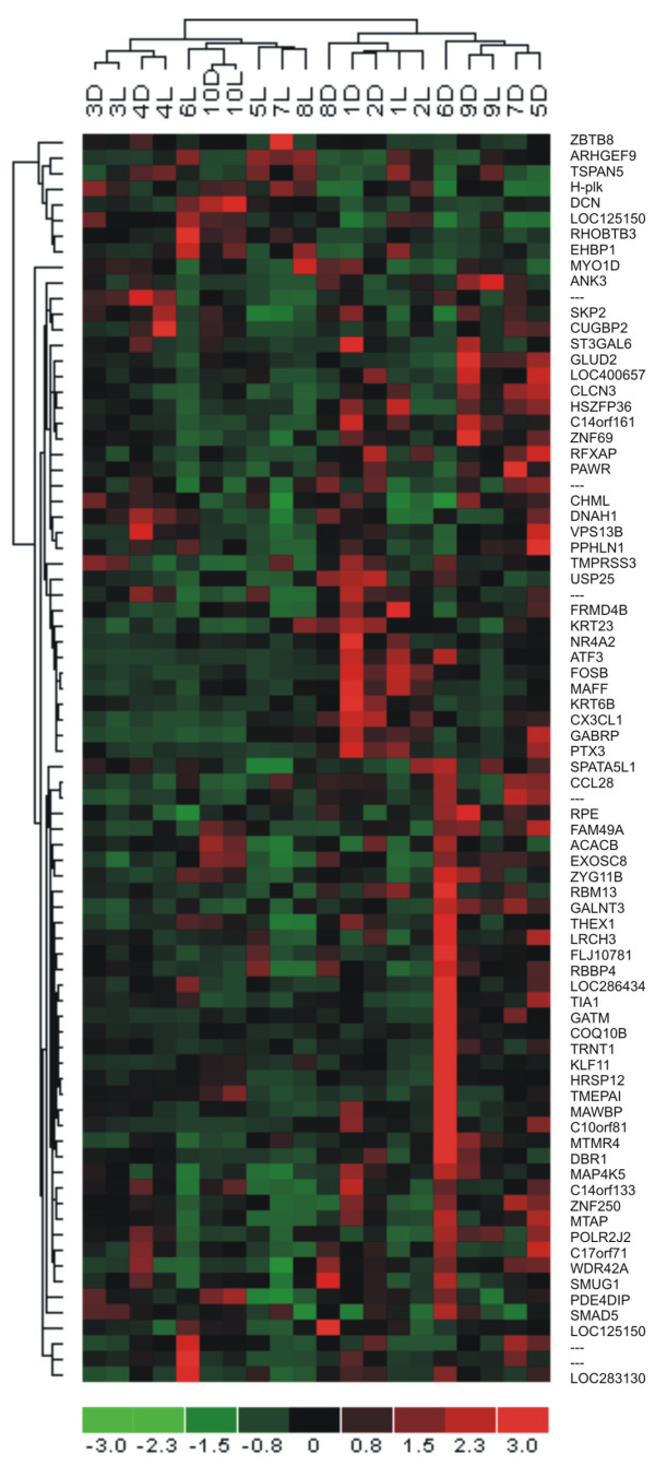
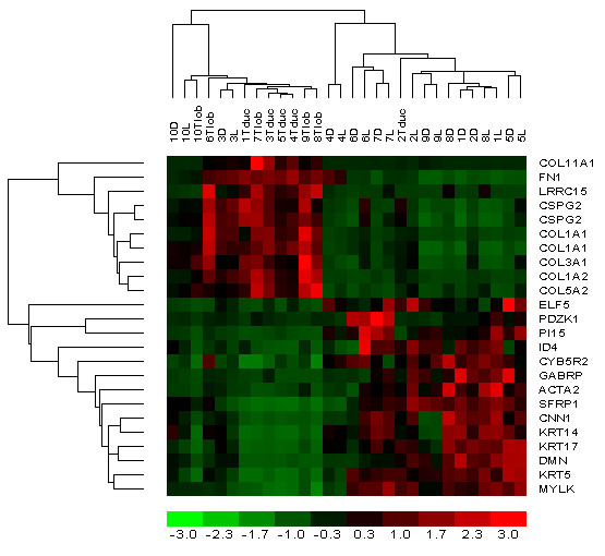
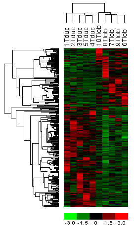
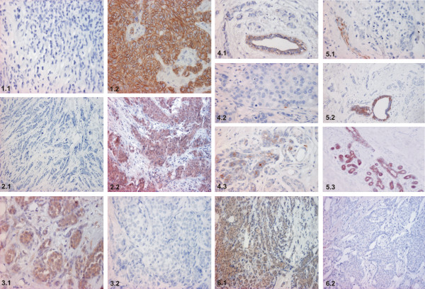
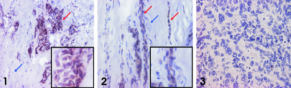
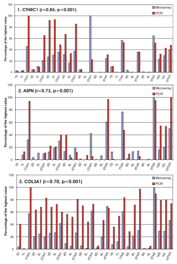
Methods: We examined 30 samples (normal ductal and lobular cells from 10 patients, IDC cells from 5 patients, ILC cells from 5 patients) microdissected from cryosections of ten mastectomy specimens from postmenopausal patients. Fifty nanograms of total RNA were amplified and labeled by PCR and in vitro transcription. Samples were analysed upon Affymetrix U133 Plus 2.0 Arrays. The expression of seven differentially expressed genes (CDH1, EMP1, DDR1, DVL1, KRT5, KRT6, KRT17) was verified by immunohistochemistry on tissue microarrays. Expression of ASPN mRNA was validated by in situ hybridization on frozen sections, and CTHRC1, ASPN and COL3A1 were tested by PCR.
Results: Using GCOS pairwise comparison algorithm and rank products we have identified 84 named genes common to ILC versus normal cell types, 74 named genes common to IDC versus normal cell types, 78 named genes differentially expressed between normal ductal and lobular cells, and 28 named genes between IDC and ILC. Genes distinguishing between IDC and ILC are involved in epithelial-mesenchymal transition, TGF-beta and Wnt signaling. These changes were present in both tumor types but appeared to be more prominent in ILC. Immunohistochemistry for several novel markers (EMP1, DVL1, DDR1) distinguished large sets of IDC from ILC.
Conclusion: IDC and ILC can be differentiated both at the gene and protein levels. In this study we report two candidate genes, asporin (ASPN) and collagen triple helix repeat containing 1 (CTHRC1) which might be significant in breast carcinogenesis. Besides E-cadherin, the proteins validated on tissue microarrays (EMP1, DVL1, DDR1) may represent novel immunohistochemical markers helpful in distinguishing between IDC and ILC. Further studies with larger sets of patients are needed to verify the gene expression profiles of various histological types of breast cancer in order to determine molecular subclassifications, prognosis and the optimum treatment strategies.
Figures
Similar articles
-
Expression of DDR1 and DVL1 in invasive ductal and lobular breast carcinoma does not correlate with histological type, grade and hormone receptor status.Asian Pac J Cancer Prev. 2015;16(6):2385-90. doi: 10.7314/apjcp.2015.16.6.2385. Asian Pac J Cancer Prev. 2015. PMID: 25824769
-
Cadherin-catenin complex dissociation in lobular neoplasia of the breast.Breast Cancer Res Treat. 2012 Apr;132(2):641-52. doi: 10.1007/s10549-011-1860-0. Epub 2011 Nov 13. Breast Cancer Res Treat. 2012. PMID: 22080244 Free PMC article.
-
Novel immunohistochemical markers for the differentiation of lobular and ductal invasive breast carcinomas.Biomed Pap Med Fac Univ Palacky Olomouc Czech Repub. 2007 Jun;151(1):59-64. doi: 10.5507/bp.2007.010. Biomed Pap Med Fac Univ Palacky Olomouc Czech Repub. 2007. PMID: 17690741
-
Comprehensive Review of Molecular Mechanisms and Clinical Features of Invasive Lobular Cancer.Oncologist. 2021 Jun;26(6):e943-e953. doi: 10.1002/onco.13734. Epub 2021 Mar 16. Oncologist. 2021. PMID: 33641217 Free PMC article. Review.
-
E-Cadherin-Mediated Cell-Cell Adhesion and Invasive Lobular Breast Cancer.Adv Exp Med Biol. 2025;1464:259-275. doi: 10.1007/978-3-031-70875-6_14. Adv Exp Med Biol. 2025. PMID: 39821030 Review.
Cited by
-
Dichotomous roles for the orphan nuclear receptor NURR1 in breast cancer.BMC Cancer. 2013 Mar 21;13:139. doi: 10.1186/1471-2407-13-139. BMC Cancer. 2013. PMID: 23517088 Free PMC article.
-
Quantitative proteomics reveals FLNC as a potential progression marker for the development of hepatocellular carcinoma.Oncotarget. 2016 Oct 18;7(42):68242-68252. doi: 10.18632/oncotarget.11921. Oncotarget. 2016. PMID: 27626164 Free PMC article.
-
Long noncoding RNA DLEU2 and ROR1 pathway induces epithelial-to-mesenchymal transition and cancer stem cells in breast cancer.Cell Death Discov. 2024 Jan 31;10(1):61. doi: 10.1038/s41420-024-01829-3. Cell Death Discov. 2024. PMID: 38296962 Free PMC article.
-
Identification of novel susceptibility markers for the risk of overall breast cancer as well as subtypes defined by hormone receptor status in the Chinese population.J Hum Genet. 2016 Dec;61(12):1027-1034. doi: 10.1038/jhg.2016.97. Epub 2016 Sep 8. J Hum Genet. 2016. PMID: 27604554
-
An integrated bioinformatics analysis of potential therapeutic targets among matrix metalloproteinases in breast cancer.Oncol Lett. 2019 Sep;18(3):2985-2994. doi: 10.3892/ol.2019.10669. Epub 2019 Jul 26. Oncol Lett. 2019. PMID: 31452777 Free PMC article.
References
-
- Rosen PP. Rosen's breast pathology. Lippincots-Raven Publishers; 1997.
-
- Weidner N, Cote R, Suster S, Weiss L. Modern Surgical Pathology. Elsevier Science; 2003.
Publication types
MeSH terms
Substances
Grants and funding
LinkOut - more resources
Full Text Sources
Other Literature Sources
Medical
Molecular Biology Databases
Research Materials
Miscellaneous
