. 2007 Mar 26;176(7):919-28.
doi: 10.1083/jcb.200609044.

Bub1 is essential for assembly of the functional inner centromere

Affiliations

Bub1 is essential for assembly of the functional inner centromere

Yekaterina Boyarchuk et al. J Cell Biol. .

Abstract

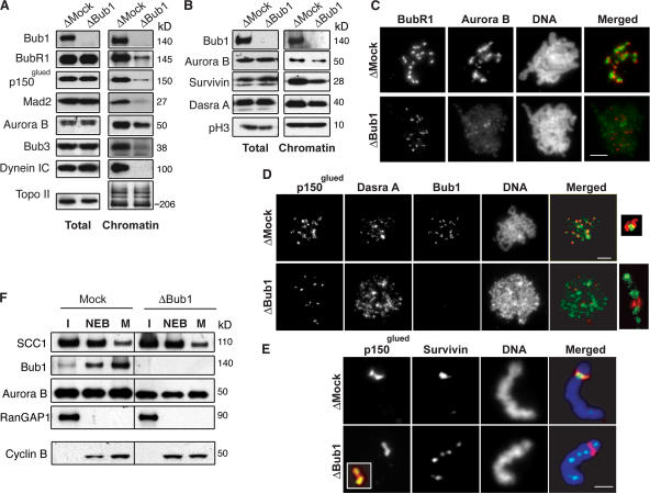
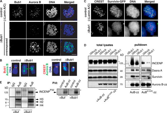
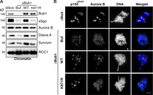
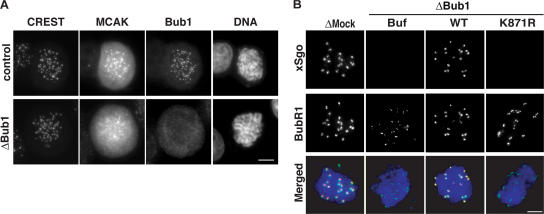
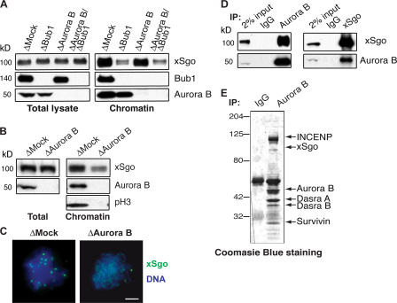
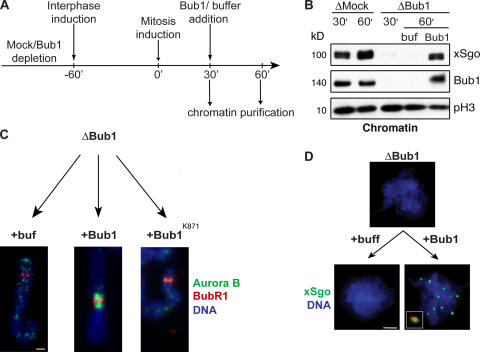
During mitosis, the inner centromeric region (ICR) recruits protein complexes that regulate sister chromatid cohesion, monitor tension, and modulate microtubule attachment. Biochemical pathways that govern formation of the inner centromere remain elusive. The kinetochore protein Bub1 was shown to promote assembly of the outer kinetochore components, such as BubR1 and CENP-F, on centromeres. Bub1 was also implicated in targeting of Shugoshin (Sgo) to the ICR. We show that Bub1 works as a master organizer of the ICR. Depletion of Bub1 from Xenopus laevis egg extract or from HeLa cells resulted in both destabilization and displacement of chromosomal passenger complex (CPC) from the ICR. Moreover, soluble Bub1 controls the binding of Sgo to chromatin, whereas the CPC restricts loading of Sgo specifically onto centromeres. We further provide evidence that Bub1 kinase activity is pivotal for recruitment of all of these components. Together, our findings demonstrate that Bub1 acts at multiple points to assure the correct kinetochore formation.


Figures

Figure 1.
Figure 1.
Bub1 controls the localization of the CPC at the ICR in X. laevis egg extracts. Mock- or Bub1-depleted CSF extracts containing 1,000 sperm/μl were driven into interphase. After 60 min, mitosis was reestablished by the addition of an aliquot of corresponding CSF-arrested extracts containing nocodazole. (A and B) Samples of total extracts or purified mitotic chromatin were analyzed by Western blotting with antibodies against indicated proteins. (C and D) Chromatin assembled as in A was purified and analyzed by indirect immunofluorescence with antibodies against Aurora B and BubR1; Dasra A and p150glued and Bub1 (unmerged). DNA was visualized with Hoechst 33342 (unmerged). Insets show magnified image of individual kinetochore pairs. In the case of Bub1-depleted extracts, note that the acquired signals for Aurora B, BubR1, and Dasra A were slightly overexposed during the processing (see the text for explanation). (E) Individual chromosomes stained with antibodies against Survivin and p150glued are shown. Inset shows magnified image of the same kinetochore stained with antibodies against CENP-A (green) and p150glued (red). (F) Chromatin assembled in mock- or Bub1-depleted extracts was purified at interphase (I, 50 min after initiation), NEB (10 min after initiation of mitosis), and metaphase (M, 30 min after initiation of mitosis), and then probed for the presence of indicated proteins by Western blotting. Antibody against RanGap1 was used to detect NEB. Total lysates of egg extracts were probed with antibody against Cyclin B to estimate stability of mitosis (bottom). Bars: (C and D) 20 μm; (E) 3 μm.
Figure 2.
Figure 2.
Bub1 is required for the inner centromeric localization of CPC in somatic cells. HeLa cells were transfected with Lamin A/C or Bub1 siRNA duplexes for 24 h and treated with nocodazole for 1 h before fixation. (A) Cells were stained to detect Bub1 (red) and Aurora B (green). (B) Individual chromosomes are shown in higher magnification to allow comparison of the Aurora B staining pattern to that of Bub1 or to the centromere marker (CREST). DNA was visualized as in Fig. 1. Note that the acquired signal of Aurora B staining in Bub1-depleted cells was overexposed. (C) HeLa cells stably expressing GFP-Survivin were treated as in A, and the position of GFP signal pattern was compared with that of CREST. (D) Bub1 is required for stability of CPC. Recombinant wtAurora B-zz or Aurora BK122R–zz were added to control or Bub1-depleted CSF extracts at a concentration approximately equal to that of endogenous Aurora B, and CPC complexes that formed on Aurora B-zz were purified by IgG–Sepharose beads. Total extracts (left) and eluates (right) were probed for the presence of CPC components. (E) Bub1 phosphorylates INCENP. CPC was either precipitated from Bub1-depleted egg extracts using antibodies against Aurora B (IP; left) or was purified on IgG–Sepharose from extracts, supplemented with Aurora B-zz (P/d, right). Mock-treated beads and beads containing precipitates were incubated with baculovirus-expressed 6His-xBub1 (50 μM) or buffer in the presence of γ[32P]ATP. Phosphate incorporation was detected using a PhosphorImager. The asterisks show the position of 6His-xBub1 (top). Immunoprecipitates (as in Fig. 1) were also incubated with core histones as exogenous substrates in the presence or absence of 6His-xBub1 to monitor Aurora B activity (bottom).
Figure 3.
Figure 3.
Bub1 kinase activity is required for ICR assembly. (A and B) Bub1-depleted extracts were supplemented either with buffer (Buf) or with in vitro–translated wild-type (WT) or kinase-dead (K871R) versions of X. laevis Bub1. (A) Mitotic chromatin assembled in these and mock-depleted extracts were analyzed for the abundance of Bub1, Sgo, and the components of the CPC by Western blotting. Antibody against RCC1 was used as a loading control. (B) Assembled chromatin was analyzed by indirect immunofluorescence with antibodies against Aurora B (green), p150glued (red), and Bub1 (blue). Bar, 20 μm.
Figure 4.
Figure 4.
Bub1 controls localization of MCAK and xSgo to the ICR. (A) Cells treated as in Fig. 2 were stained with antibodies against Bub1, MCAK, and CREST. (B) Chromatin assembled as in Fig. 3 was analyzed by indirect immunofluorescence with antibodies against xSgo (red) and anti-BubR1 (green). DNA was stained as in A (blue).
Figure 5.
Figure 5.
Soluble Bub1 is able to promote xSgo binding to the mitotic chromatin, whereas the CPC directs xSgo to the ICR. (A) Total extracts and mitotic chromatin purified from control extracts or extracts lacking Bub1, Aurora B, or both Bub1 and Aurora B, were analyzed by Western blotting with indicating antibodies. (B–E) Mock- or Aurora B-depleted extracts were treated as in Fig. 1 A and analyzed by Western blotting with the indicated antibodies (B) or by immunofluorescence with antibodies against xSgo (C; green). DNA, stained as in Fig. 1, is shown in blue. (D) Aurora B and xSgo were immunoprecipitated from CSF-arrested egg extracts by the corresponding antibodies. Immunoprecipitates were subjected to Western blotting with antibodies against Aurora B and xSgo. (E) Chromosome passenger complex precipitated with anti–Aurora B antibodies was analyzed by SDS-PAGE and CBB staining. Arrows indicate the positions of the CPC components and xSgo.
Figure 6.
Figure 6.
The assembly of the ICR is independent of mitotic chromosome formation. (A) Schematic overview of the experiment. Mock- or Bub1-depleted extracts were driven into interphase. After 60 min, mitosis was reestablished by addition of an aliquot of mock- or Bub1-depleted CSF-arrested extracts together with nocodazole. 30 min after induction of mitosis, baculovirus-expressed 6His-tagged wtBub1, Bub1K871R, or buffer were added, followed by incubation for an additional 30 min. (B) Chromatin was analyzed by Western blotting for the abundance of xSgo, Bub1, and phosphorylated histone H3 (pH3). (C and D) Purified chromatin was stained with antibody against Aurora B (C, green) and BubR1 (C, red) or xSgo (D, green). DNA was visualized as in Fig. 1 (blue). The inset in D shows a magnified image of an individual kinetochore pair stained with antibody against Bub1 (red) and xSgo (green). Bars: (C) 2 μm; (D) 20 μm.

References

    1. Andrews, P.D., E. Knatko, W.J. Moore, and J.R. Swedlow. 2003. Mitotic mechanics: the auroras come into view. Curr. Opin. Cell Biol. 15:672–683. - PubMed
    1. Andrews, P.D., Y. Ovechkina, N. Morrice, M. Wagenbach, K. Duncan, L. Wordeman, and J.R. Swedlow. 2004. Aurora B regulates MCAK at the mitotic centromere. Dev. Cell. 6:253–268. - PubMed
    1. Azuma, Y., A. Arnaoutov, and M. Dasso. 2003. SUMO-2/3 regulates topoisomerase II in mitosis. J. Cell Biol. 163:477–487. - PMC - PubMed
    1. Azuma, Y., A. Arnaoutov, T. Anan, and M. Dasso. 2005. PIASy mediates SUMO-2 conjugation of topoisomerase-II on mitotic chromosomes. EMBO J. 24:2172–2182. - PMC - PubMed
    1. Chan, G.K., S.T. Liu, and T.J. Yen. 2005. Kinetochore structure and function. Trends Cell Biol. 15:589–598. - PubMed

Publication types

MeSH terms