. 2007 Mar 26;176(7):941-52.
doi: 10.1083/jcb.200701048.

Jagunal is required for reorganizing the endoplasmic reticulum during Drosophila oogenesis

Affiliations

Jagunal is required for reorganizing the endoplasmic reticulum during Drosophila oogenesis

Sangil Lee et al. J Cell Biol. .

Abstract

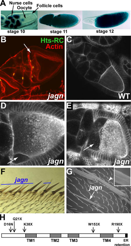
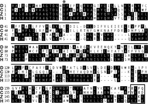
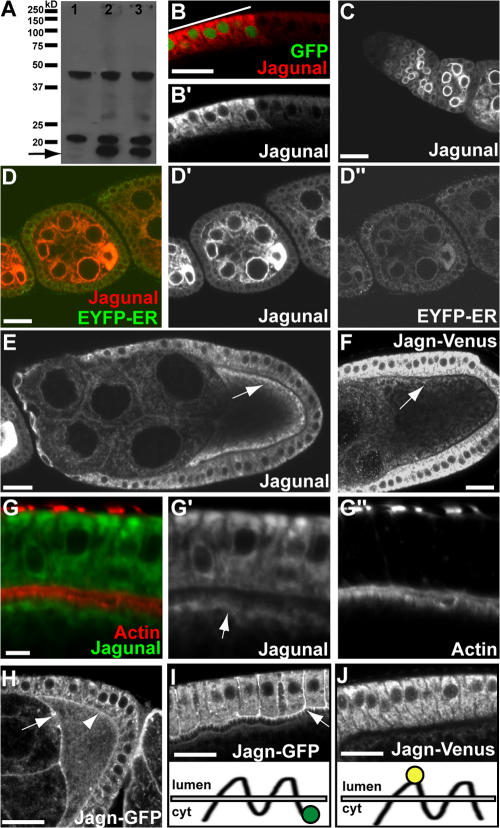
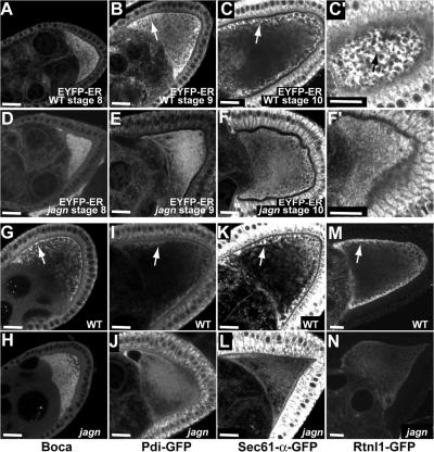
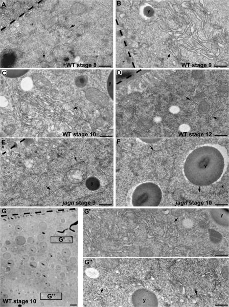
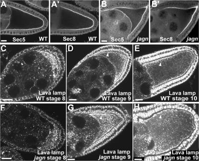
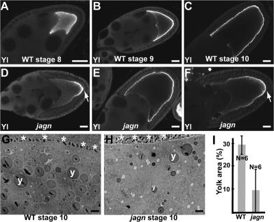
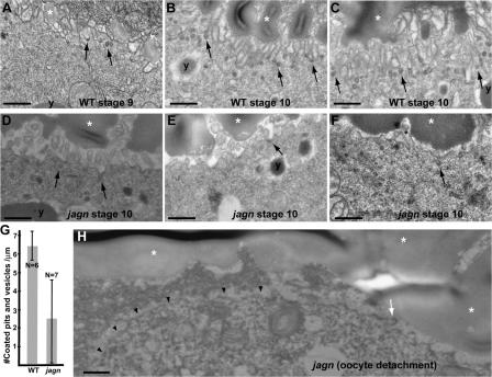
Vesicular traffic in the Drosophila melanogaster oocyte occurs actively during vitellogenesis. Although endocytosis in the oocyte has been well characterized, exocytic vesicular traffic is less well understood. We show that the oocyte endoplasmic reticulum (ER) becomes concentrated into subcortical clusters during vitellogenesis. This ER reorganization requires Jagunal, which is an evolutionarily conserved ER membrane protein. Loss of Jagunal reduces vesicular traffic to the oocyte lateral membrane, but does not affect posterior polarized vesicular traffic, suggesting a role for Jagunal in facilitating vesicular traffic in the subcortex. Reduced membrane traffic caused by loss of Jagunal affects oocyte and bristle growth. We propose that ER reorganization is an important mechanism used by cells to prepare for an increased demand for membrane traffic, and Jagunal facilitates this process through ER clustering.


Figures

Figure 1.
Figure 1.
Jagunal is required for oocyte and bristle growth. (A) D. melanogaster egg chambers. Egg chambers before (stage 10), during (stage 11), and after (stage 12) nurse-cell dumping are shown with nuclear lacZ staining. Egg chambers are composed of germline cells (15 nurse cells and the oocyte) surrounded by somatic follicle cells. All egg chamber images are oriented with anterior to the left. (B) jagnQ21X GLCs were stained with rhodamine-conjugated phalloidin (red) to visualize F-actin and Hts-RC antibodies (green) to visualize ring canals. Mutant oocytes detach from nurse cells and follicle cells at the anterior region (arrow). (C–E) Live egg chambers were examined by Bsg-GFP fluorescence, which reveals plasma membranes and vesicles throughout the ooplasm. Wild-type egg chambers (C) and jagnQ21X GLCs (D and E) are shown. Mutant oocytes detach from nurse cells and follicle cells at the anterior region (D, arrow), and the defect becomes more severe as oogenesis proceeds (E, arrow). (F) jagnQ21X clones were marked by yellow (y ). Mutant wing anterior margin bristles are thinner and shorter than their wild-type counterparts. (G) A scanning electron microscope image of wild-type (arrowhead) and jagnQ21X (arrow) microchaetes. Mutant microchaetes are thinner and shorter than wild-type microchaetes, and have weak ridges (inset in G). (H) Domains of Jagunal and mutation sites in jagn alleles. Jagunal contains four predicted transmembrane domains (TMs) and a putative ER retention motif (dilysine motif) at the C-terminal end. Mutation sites in all jagn alleles were identified in the open reading frame of the jagunal gene (CG10978). jagnQ21X is the original mutation isolated from the Lehmann collection, and the other four alleles were isolated from a noncomplementation lethal screen.
Figure 2.
Figure 2.
Alignment of Jagunal homologues. Jagunal homologues from D. melanogaster (NP_649585; D), C. elegans (NP_493559; C), zebrafish (NP_001005774; Z), and human (AAH32101; H) are shown. Four predicted transmembrane domains in D. melanogaster Jagunal are indicated by lines drawn over the sequence, and a putative dilysine motif at the C-terminal end is indicated by a boxed area. The mutation site in a semilethal allele, jagnD16N, is indicated by an asterisk.
Figure 3.
Figure 3.
Localization of Jagunal in egg chambers. (A) Western blot analysis of wild-type (lane 1) and Jagunal-overexpressing (lanes 2 and 3) ovary extracts. The actin-Gal4 driver (lane 2) and germline triple drivers (lane 3) were used to overexpress Jagunal in the ovary. In wild-type extract, two nonspecific bands were detected, but endogenous Jagunal was barely visible. However, overexpressed Jagunal was detected (arrow). (B and B′) Jagunal-overexpressing follicle cells were marked with GFP (indicated by a bar). Jagunal expression was elevated in GFP-positive cells (red). The follicle cells are shown with the basal membrane at the top. (C–E and G) Jagunal was overexpressed using the actin-Gal4 driver. (C) Jagunal is enriched at the nuclear envelope during early stages. (D–D″) Egg chambers expressing EYFP-ER (green) were stained for Jagunal (red). Jagunal colocalizes with EYFP-ER and becomes enriched in the oocyte during early stages. (E and G) Jagunal becomes enriched at the oocyte subcortex during stages 9 and 10 (arrows). (G–G″) Jagunal enriched at the oocyte subcortex (arrow) is near cortical actin, but does not colocalize with cortical actin. (F and J) Localization of Jagn-Venus in egg chambers. For the Jagn-Venus construct, Venus was inserted between the first and second transmembrane domains (J, bottom). Jagn-Venus localizes at the ER. (H and I) Localization of Jagn-GFP in egg chambers. For the Jagn-GFP construct, GFP was fused to the C terminus (I, bottom). In addition to ER localization, Jagn-GFP localizes at the plasma membrane of follicle cells (I, arrow) and the oocyte (H, arrowhead), including ring canals (H, arrow). Localization of Jagn-GFP to follicle cell plasma membrane is specific to stage 10. Bars: (A–F and H–J) 20 μm; (G) 5 μm.
Figure 4.
Figure 4.
Jagunal is required for the enrichment of ER proteins in the oocyte subcortical region during vitellogenesis. (A–F) The distribution of EYFP-ER in progressive stages of wild-type egg chambers (A–C) and jagnQ21X GLCs (D–F). Until stage 8, EYFP-ER distributes uniformly in wild-type oocytes (A). However, EYFP-ER becomes enriched in the oocyte subcortex during stages 9 and 10 (B and C, arrows). (C′) A focal plane near the cortex of the oocyte shown in C shows that EYFP-ER is concentrated into clusters in the oocyte subcortex (arrow). (D–F) Until stage 8, EYFP-ER distributes normally in jagn mutant oocytes (D). However, EYFP-ER remains dispersed in jagn mutant oocytes, with very little subcortical enrichment during stages 9 and 10 (E and F). (F′) A focal plane near the cortex of the oocyte shown in F shows that EYFP-ER distributes uniformly without forming clusters in the oocyte subcortex. (G–N) The distribution of ER proteins Boca (G and H), PDI-GFP (I and J), Sec61-α-GFP (K and L), and Rtnl1-GFP (M and N) in wild-type egg chambers and jagnQ21X GLCs. Similar to EYFP-ER, these ER proteins become enriched in the subcortical region in wild-type oocytes (G, I, K, and M, arrows). The enrichment of these proteins in the subcortex is reduced in jagn mutant oocytes (H, J, L, and N). Bars, 20 μm.
Figure 5.
Figure 5.
Jagunal is required for ER clustering in the oocyte during vitellogenesis. (A–D) EM images of progressive stages of wild-type egg chambers. During stage 8, the ER density is low and ER clusters are not found in the oocyte (A). ER clusters are formed in the oocyte, and the ER luminal space increases during stages 9 and 10 (B and C). ER clusters are not found in the oocyte during stage 12 (D). (E and F) EM images of stage 9 and 10 jagnQ21X GLCs. ER clusters are not found in jagn mutant oocytes, and the ER luminal space does not increase during stages 9 and 10 (E and F). (G) EM image of stage 10 oocyte. The ER density in the subcortical region (G′) is higher than inside of the ooplasm (G″). (A–G) Oocyte–follicle cell boundaries and yolk granules are marked by dashed lines and y's, respectively. Arrows indicate the ER. Bars: (A–F, G′, and G″) 0.5 μm; (G) 2 μm.
Figure 6.
Figure 6.
Exocyst and Golgi complexes distribute normally in jagn mutant oocytes. (A and B) Two exocyst components, Sec5 and Sec8, were examined in wild-type egg chambers (A) and jagnQ21X GLCs (B). In mutant oocytes, Sec5 and Sec8 localize normally at the cortex (B). Progressive stages of wild-type egg chambers (C–E) and jagnQ21X GLCs (F–H) were stained with Lava lamp antibodies to detect Golgi complexes. In wild-type stage 8 egg chambers, Golgi complexes are enriched in the oocyte and distributed evenly in the oocyte cytoplasm (C). During stages 9 and 10, many Golgi complexes accumulate near the oocyte cortex (D and E, arrowheads). In mutant oocytes, Golgi complex distribution is normal, with many Golgi complexes enriched in the subcortex (G and H, arrowhead). Bars, 20 μm.
Figure 7.
Figure 7.
Transport of Yolkless to the oocyte lateral membrane is reduced in jagn GLCs. (A–F) Wild-type egg chambers (A–C) and jagnQ21X GLCs (D–F) were stained with Yolkless antibodies. Yolkless begins to be transported to the oocyte surface during stage 8 (A). During stages 9 and 10, Yolkless is localized at the oocyte cortex adjacent to follicle cells (B and C). In mutant oocytes (D–F), the intensity of Yolkless staining at the oocyte cortex is reduced. In most egg chambers the distribution of Yolkless is uneven, with the highest level at the posterior region (D and F, arrows). Yolkless enrichment at the posterior region is most evident during stage 9 (D). (G and H) EM images of a wild-type egg chamber (G) and a jagnQ21X GLC (H). Asterisks and y's mark oocyte–follicle cell boundary and yolk granules, respectively. (I) Quantitation of the percentage of yolk area in wild-type egg chambers and jagnQ21X GLCs. Percentage of yolk area was defined as the percentage of area occupied by yolk granules in the ooplasm. N indicates the number of examined oocytes. Bars: (A–F) 20 μm; (G and H) 2 μm.
Figure 8.
Figure 8.
Endocytosis and cell surface area is reduced in jagn mutant oocytes. EM images of progressive stages of wild-type egg chambers (A–C) and stage 10 jagnQ21X GLCs (D–F). Arrows indicate coated pits and vesicles. y's and asterisks mark yolk granules and vitelline bodies, respectively. In wild-type egg chambers, the oocyte surface has a high density of microvilli, and many coated pits and vesicles are found in the plasma membrane and cortex, especially during stage 10 (A–C). In jagn mutant oocytes, the density of microvilli is reduced to varying degrees, resulting in a decrease of the overall cell surface area (D–F). The number of coated pits and vesicles are also reduced in jagn mutant oocytes (D–F). (G) Quantitation of the number of coated pits and vesicles/micrometer. The number of coated pits and vesicles was counted and divided by the linear length of the examined oocyte surface. N indicates the number of examined oocytes. (H) An EM image of the anterior region of jagnQ21X mutant oocyte. The plasma membrane of the mutant oocyte (arrowheads) is detached from neighboring cells. The number of microvilli (indicated by an arrow) is reduced. Bars, 0.5 μm.

Similar articles

Cited by

References

    1. Ashburner, M. 1989. Drosophila: A Laboratory Handbook. Cold Spring Harbor Laboratory Press, Cold Spring Harbor, New York.
    1. Baumann, O., and B. Walz. 2001. Endoplasmic reticulum of animal cells and its organization into structural and functional domains. Int. Rev. Cytol. 205:149–214. - PubMed
    1. Beronja, S., P. Laprise, O. Papoulas, M. Pellikka, J. Sisson, and U. Tepass. 2005. Essential function of Drosophila Sec6 in apical exocytosis of epithelial photoreceptor cells. J. Cell Biol. 169:635–646. - PMC - PubMed
    1. Bobinnec, Y., C. Marcaillou, X. Morin, and A. Debec. 2003. Dynamics of the endoplasmic reticulum during early development of Drosophila melanogaster. Cell Motil. Cytoskeleton. 54:217–225. - PubMed
    1. Bretscher, M.S. 1996. Expression and changing distribution of the human transferrin receptor in developing Drosophila oocytes and embryos. J. Cell Sci. 109:3113–3119. - PubMed

Publication types

MeSH terms