. 2007 Apr 3;104(14):5800-5.
doi: 10.1073/pnas.0700979104. Epub 2007 Mar 26.

Interdomain communication in calcium pump as revealed in the crystal structures with transmembrane inhibitors

Affiliations

Interdomain communication in calcium pump as revealed in the crystal structures with transmembrane inhibitors

Mihoko Takahashi et al. Proc Natl Acad Sci U S A. .

Abstract

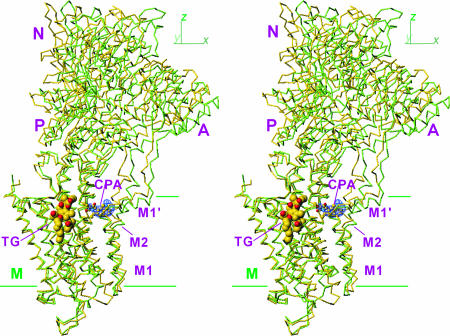
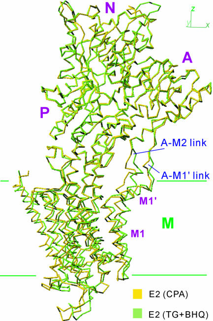
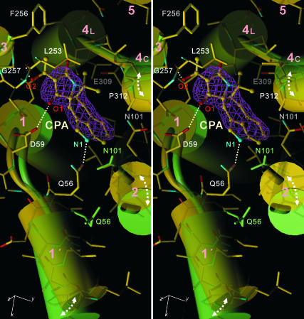
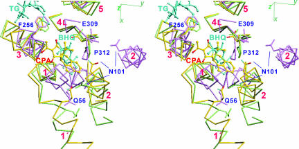
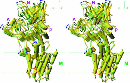
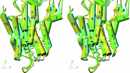
Ca(2+)-ATPase of skeletal muscle sarcoplasmic reticulum is an ATP-driven Ca(2+) pump consisting of three cytoplasmic domains and 10 transmembrane helices. In the absence of Ca(2+), the three cytoplasmic domains gather to form a compact headpiece, but the ATPase is unstable without an inhibitor. Here we describe the crystal structures of Ca(2+)-ATPase in the absence of Ca(2+) stabilized with cyclopiazonic acid alone and in combination with other inhibitors. Cyclopiazonic acid is located in the transmembrane region of the protein near the cytoplasmic surface. The binding site partially overlaps with that of 2,5-di-tert-butyl-1,4-dihydroxybenzene but is separate from that of thapsigargin. The overall structure is significantly different from that stabilized with thapsigargin: The cytoplasmic headpiece is more upright, and the transmembrane helices M1-M4 are rearranged. Cyclopiazonic acid primarily alters the position of the M1' helix and thereby M2 and M4 and then M5. Because M5 is integrated into the phosphorylation domain, the whole cytoplasmic headpiece moves. These structural changes show how an event in the transmembrane domain can be transmitted to the cytoplasmic domain despite flexible links between them. They also reveal that Ca(2+)-ATPase has considerable plasticity even when fixed by a transmembrane inhibitor, presumably to accommodate thermal fluctuations.


Conflict of interest statement

The authors declare no conflict of interest.

Figures

Fig. 1.
Fig. 1.
Superimposition of E2(CPA) and E2(TG+BHQ), which represent the two most distant structures in the five E2 crystals. Cα traces are viewed in stereo along the plane of the lipid bilayer (i.e., xy-plane). Two structures are fitted with the M7–M10 transmembrane helices. Yellow, E2(CPA); light green, E2(TG+BHQ). TG is shown in space fill, and CPA is in ball-and-stick. The blue net shows an ∣Fobs∣ − ∣Fcalc∣ electron density map (contoured at 5σ) before introduction of CPA in the atomic model. Three cytoplasmic (A, N, and P) and transmembrane (M) domains are marked.
Fig. 2.
Fig. 2.
Superimposition using the P-domain showing that the headpiece moves together and that the links between the A-domain and transmembrane helices M1–M2 are flexible. The structures shown here are viewed in the direction shown in Fig. 1.
Fig. 3.
Fig. 3.
The CPA binding site in E2(CPA+CC) superimposed with that of E2(TG+BHQ) viewed in stereo approximately perpendicular to the membrane. Yellow, E2(CPA+CC); light green, E2(TG+BHQ). Cylinders represent transmembrane helices (M1–M5). The pink net represents an ∣Fobs∣ − ∣Fcalc∣ electron density map at 2.8-Å resolution contoured at 4σ before CPA (shown in ball-and-stick) was introduced into the atomic model. Dotted lines show likely hydrogen bonds. The atomic model is shown in stick representation for E2(CPA+CC). Side chains of Asn-101 and Gln-56 also are shown for E2(TG+BHQ).
Fig. 4.
Fig. 4.
Three transmembrane inhibitors (CPA, TG, and BHQ) superimposed with Cα traces of the three crystal structures, E1·2Ca2+ (violet), E2(CPA+CC) (yellow), and E2(TG+BHQ) (light green), viewed in stereo. The side chains of several key residues are shown. The transmembrane helices (M1–M6) are numbered.
Fig. 5.
Fig. 5.
Global structural changes induced by the binding of CPA. E2(CPA+CC) (yellow) and E2(TG+BHQ) (light green) crystal structures are superimposed with the M7–M10 helices. The structures are shown from the side opposite to that in Fig. 1. Arrows indicate the differences between the two structures. The circle indicates the amphipathic helix between M8 and M9. CPA is shown in space fill. M2, M5, and M7 are represented with two to three cylinders, although they are continuous helices.
Fig. 6.
Fig. 6.
Differences in orientation of the transmembrane helices in three crystal structures. E2(CPA) (yellow), E2(TG+BHQ) (light green), and E2·MgF42− (cyan) are superimposed with the M7–M10 helices and are shown in stereo in the same direction as in Fig. 1. M2, M3, M5, and M7 are represented with two to three cylinders, although they are continuous helices. Some of the residues used in helix–helix contacts are shown.

References

    1. Møller JV, Juul B, le Maire M. Biochim Biophys Acta. 1996;1286:1–51. - PubMed
    1. Toyoshima C, Inesi G. Annu Rev Biochem. 2004;73:269–292. - PubMed
    1. Toyoshima C, Nakasako M, Nomura H, Ogawa H. Nature. 2000;405:647–655. - PubMed
    1. Toyoshima C, Nomura H. Nature. 2002;418:605–611. - PubMed
    1. Toyoshima C, Mizutani T. Nature. 2004;430:529–535. - PubMed

Publication types

MeSH terms

LinkOut - more resources