. 2007 Apr 3;104(14):6031-6.
doi: 10.1073/pnas.0700036104. Epub 2007 Mar 26.

Lysosomal killing of Mycobacterium mediated by ubiquitin-derived peptides is enhanced by autophagy

Affiliations

Lysosomal killing of Mycobacterium mediated by ubiquitin-derived peptides is enhanced by autophagy

Sylvie Alonso et al. Proc Natl Acad Sci U S A. .

Abstract

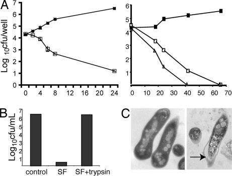
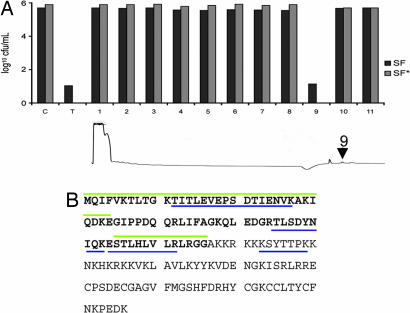
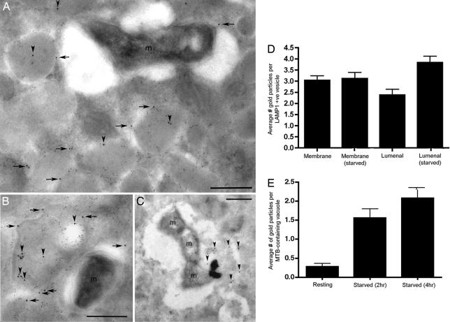
Mycobacterium tuberculosis parasitizes resting macrophages yet is killed by activated macrophages through both oxidative and nonoxidative mechanisms. Nonoxidative mechanisms are linked to the maturation of the bacteria-containing phagosome into an acidified, hydrolytically active compartment. We describe here a mechanism for killing Mycobacteria in the lysosomal compartment through the activity of peptides generated by the hydrolysis of ubiquitin. The induction of autophagy in infected macrophages enhanced the delivery of ubiquitin conjugates to the lysosome and increased the bactericidal capacity of the lysosomal soluble fraction. The accumulation of ubiquitinated proteins in the autophagolysosome provides one possible mechanism behind the antimicrobial activities observed for a range of pathogens in autophagous host cells.


Conflict of interest statement

The authors declare no conflict of interest.

Figures

Fig. 1.
Fig. 1.
Bactericidal activity of lysosomal SF on M. smegmatis and M. tuberculosis. (A) Bacteria were incubated with buffer (filled squares) or with SF at 50 μg/ml (open squares) or 100 μg/ml (triangles). At the indicated time points, the viable bacteria were determined by plating cfu. (Left) M. smegmatis. (Right) M. tuberculosis. (B) The bactericidal activity of lysosomal SF is protease-sensitive. M. smegmatis was incubated overnight at 37°C with buffer, SF, or SF preincubated with trypsin, then the viable bacteria were determined. (C) SF-treated mycobacteria display membrane damage. M. smegmatis was incubated overnight with buffer or SF and then analyzed by electron microscopy. (Left) Control. (Right) SF-treated. An arrow indicates the compromised cell wall.
Fig. 2.
Fig. 2.
Identification of ubiquitin peptides by mass spectrometry. (A) HPLC purification of the fraction containing mycobactericidal compound. The bactericidal activity of the total (T) sample was compared with that of the buffer-treated control (C) and each fraction from SF and SF*. The arrow on the trace indicates the peak retaining bactericidal activity. (B) Amino acid sequence of ubiquitin as an N-terminal extension of ribosomal subunit Rps27A. Amino acids corresponding to the ubiquitin are in bold. The peptides identified by mass spectroscopy are underlined in blue, and the peptides Ub1 and Ub2 are overlined in green.
Fig. 3.
Fig. 3.
Immunoelectron microscopy of M. tuberculosis-infected macrophages. Cells were probed with mouse antiubiquitin (12 nm gold) and rat anti-LAMP1 (6 nm gold). (A) Untreated, infected macrophage demonstrating that the bacteria-containing vacuoles have minimal ubiquitin signal. The ubiquitin signal is associated predominantly with the limiting membranes (horizontal arrows) or lumen (vertical arrowheads) of LAMP1-positive vesicles. (B) Untreated, infected macrophages where bacteria-containing vacuoles have minimal ubiquitin signal and where the ubiquitin signal is associated predominantly with internal, LAMP1-positive membranous material inside MVBs (vertical arrowheads). (C) In cells treated by starvation for 2 h, M. tuberculosis can be seen in vacuoles with flocculent lysosomal matrix that is also positive for ubiquitin (vertical arrowheads). (D) The distribution of ubiquitin signal associated with the limiting membrane or the lumen of LAMP1-positive vesicles was scored in resting versus 2-h starved macrophages and showed a significant increase in luminal label in the starved cells (n = 50 vacuoles). (E) The relative distribution of M. tuberculosis in vacuoles positive for luminal ubiquitin signal in untreated and starved BMMΦ is detailed ± SD (n = 50 vacuoles).
Fig. 4.
Fig. 4.
Bacterial killing by BMMΦ upon induction of autophagy and activity of SF, SF*, and SF IFN against Mycobacteria. (A) Bactericidal activity of SF and SF*. M. smegmatis (Left) and M. tuberculosis (Right) were incubated in the presence of 50 μg/ml SF or SF* purified from control and autophagic BMMΦ, and bacterial viability was determined. Statistical significance between SF and SF* treatment was determined by Student's t test: ∗, P < 0.5; ∗∗, P < 0.05; ∗∗∗, P < 0.01; ∗∗∗∗, P < 0.005. (B) Bactericidal activity against M. smegmatis of 50 μg/ml SF purified from control, SF*, and SF IFN was determined. Statistical significance between SF and SF* treatment was determined by Student's t test: ∗, P < 0.5; ∗∗, P < 0.05. (A and B) The average ± SD of three independent experiments is shown. (C) (Left) Immunoblot analysis of SF, SF*, and SF IFN compared with ubiquitin standards. (Right) Signals quantitated by densitometry.
Fig. 5.
Fig. 5.
Bactericidal activity of ubiquitin and ubiquitin-derived peptides. (A) Bactericidal activity against M. smegmatis of purified ubiquitin and cathepsin-treated ubiquitin. Bacteria were incubated with 25 μM purified ubiquitin or 25 μM preincubated with cathepsin B, D, or L overnight. (B) Bactericidal activity against M. smegmatis of purified ubiquitin and SF-treated ubiquitin. Bacteria were incubated overnight with 100 μM purified ubiquitin or 25 μM preincubated with 25 μg/ml SF (SF25). Statistical significance between SF25 and SF25+Ub100 treatment was determined by Student's t test (∗∗∗∗, P < 0.005). (C) M. tuberculosis viability in the presence of varying concentrations of Ub2 peptide over the indicated time course was determined by plating cfu.

Comment in

References

    1. Deretic V, Singh S, Master S, Harris J, Roberts E, Kyei G, Davis A, de Haro S, Naylor J, Lee HH, Vergne I. Cell Microbiol. 2006;8:719–727. - PubMed
    1. Russell DG. Nat Rev Mol Cell Biol. 2001;2:569–577. - PubMed
    1. Sturgill-Koszycki S, Schlesinger PH, Chakraborty P, Haddix PL, Collins HL, Fok AK, Allen RD, Gluck SL, Heuser J, Russell DG. Science. 1994;263:678–681. - PubMed
    1. Nathan C, Shiloh MU. Proc Natl Acad Sci USA. 2000;97:8841–8848. - PMC - PubMed
    1. MacMicking JD, Taylor GA, McKinney JD. Science. 2003;302:654–659. - PubMed

Publication types

MeSH terms