Coordinate expression and functional profiling identify an extracellular proteolytic signaling pathway
- PMID: 17389401
- PMCID: PMC1838401
- DOI: 10.1073/pnas.0606514104
Coordinate expression and functional profiling identify an extracellular proteolytic signaling pathway
Abstract
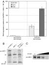
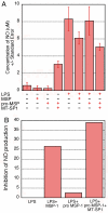
A multidisciplinary method combining transcriptional data, specificity profiling, and biological characterization of an enzyme may be used to predict novel substrates. By integrating protease substrate profiling with microarray gene coexpression data from nearly 2,000 human normal and cancerous tissue samples, three fundamental components of a protease-activated signaling pathway were identified. We find that MT-SP1 mediates extracellular signaling by regulating the local activation of the prometastatic growth factor MSP-1. We demonstrate MT-SP1 expression in peritoneal macrophages, and biochemical methods confirm the ability of MT-SP1 to cleave and activate pro-MSP-1 in vitro and in a cellular context. MT-SP1 induced the ability of MSP-1 to inhibit nitric oxide production in bone marrow macrophages. Addition of HAI-1 or an MT-SP1-specific antibody inhibitor blocked the proteolytic activation of MSP-1 at the cell surface of peritoneal macrophages. Taken together, our work indicates that MT-SP1 is sufficient for MSP-1 activation and that MT-SP1, MSP-1, and the previously shown MSP-1 tyrosine kinase receptor RON are required for peritoneal macrophage activation. This work shows that this triad of growth factor, growth factor activator protease, and growth factor receptor is a protease-activated signaling pathway. Individually, MT-SP1 and RON overexpression have been implicated in cancer progression and metastasis. Transcriptional coexpression of these genes suggests that this signaling pathway may be involved in several human cancers.
Conflict of interest statement
The authors declare no conflict of interest.
Figures


Similar articles
-
Proteolytic activation of pro-macrophage-stimulating protein by hepsin.Mol Cancer Res. 2011 Sep;9(9):1175-86. doi: 10.1158/1541-7786.MCR-11-0004. Epub 2011 Aug 29. Mol Cancer Res. 2011. PMID: 21875933
-
Activation of the stem cell-derived tyrosine kinase/RON receptor tyrosine kinase by macrophage-stimulating protein results in the induction of arginase activity in murine peritoneal macrophages.J Immunol. 2002 Jan 15;168(2):853-60. doi: 10.4049/jimmunol.168.2.853. J Immunol. 2002. PMID: 11777982
-
Hepatocyte growth factor activator is a serum activator of single-chain precursor macrophage-stimulating protein.FEBS J. 2009 Jul;276(13):3481-90. doi: 10.1111/j.1742-4658.2009.07070.x. Epub 2009 May 18. FEBS J. 2009. PMID: 19456860
-
Hepatocyte growth factor activator inhibitor type‑1 in cancer: advances and perspectives (Review).Mol Med Rep. 2014 Dec;10(6):2779-85. doi: 10.3892/mmr.2014.2628. Epub 2014 Oct 13. Mol Med Rep. 2014. PMID: 25310042 Review.
-
Kinases involved in MSP/RON signaling.J Leukoc Biol. 1999 Mar;65(3):345-8. doi: 10.1002/jlb.65.3.345. J Leukoc Biol. 1999. PMID: 10080538 Review.
Cited by
-
Impact of suppression of tumorigenicity 14 (ST14)/serine protease 14 (Prss14) expression analysis on the prognosis and management of estrogen receptor negative breast cancer.Oncotarget. 2016 Jun 7;7(23):34643-63. doi: 10.18632/oncotarget.9155. Oncotarget. 2016. PMID: 27167193 Free PMC article.
-
The cutting edge: membrane-anchored serine protease activities in the pericellular microenvironment.Biochem J. 2010 Jun 15;428(3):325-46. doi: 10.1042/BJ20100046. Biochem J. 2010. PMID: 20507279 Free PMC article. Review.
-
Polarized epithelial cells secrete matriptase as a consequence of zymogen activation and HAI-1-mediated inhibition.Am J Physiol Cell Physiol. 2009 Aug;297(2):C459-70. doi: 10.1152/ajpcell.00201.2009. Epub 2009 Jun 17. Am J Physiol Cell Physiol. 2009. PMID: 19535514 Free PMC article.
-
RON kinase inhibition reduces renal endothelial injury in sickle cell disease mice.Haematologica. 2018 May;103(5):787-798. doi: 10.3324/haematol.2017.180992. Epub 2018 Mar 8. Haematologica. 2018. PMID: 29519868 Free PMC article.
-
Epithelial integrity is maintained by a matriptase-dependent proteolytic pathway.Am J Pathol. 2009 Oct;175(4):1453-63. doi: 10.2353/ajpath.2009.090240. Epub 2009 Aug 28. Am J Pathol. 2009. PMID: 19717635 Free PMC article.
References
-
- Cao J, Zheng S, Zheng L, Cai X, Zhang Y, Geng L, Fang Y. Chin Med J (English) 2001;114:726–730. - PubMed
-
- Galkin AV, Mullen L, Fox WD, Brown J, Duncan D, Moreno O, Madison EL, Agus DB. Prostate. 2004;61:228–235. - PubMed
-
- Hoang CD, D'Cunha J, Kratzke MG, Casmey CE, Frizelle SP, Maddaus MA, Kratzke RA. Chest. 2004;125:1843–1852. - PubMed
-
- Johnson MD, Oberst MD, Lin CY, Dickson RB. Exp Rev Mol Diagn. 2003;3:331–338. - PubMed
-
- Kang JY, Dolled-Filhart M, Ocal IT, Singh B, Lin CY, Dickson RB, Rimm DL, Camp RL. Cancer Res. 2003;63:1101–1105. - PubMed
Publication types
MeSH terms
Substances
LinkOut - more resources
Full Text Sources
Other Literature Sources
Research Materials
Miscellaneous

