Comparative Study
. 2007 Apr 10;104(15):6217-22.
doi: 10.1073/pnas.0701297104. Epub 2007 Mar 27.

Structural insights into the degradation of Mcl-1 induced by BH3 domains

Affiliations
Comparative Study

Structural insights into the degradation of Mcl-1 induced by BH3 domains

Peter E Czabotar et al. Proc Natl Acad Sci U S A. .

Abstract

Apoptosis is held in check by prosurvival proteins of the Bcl-2 family. The distantly related BH3-only proteins bind to and antagonize them, thereby promoting apoptosis. Whereas binding of the BH3-only protein Noxa to prosurvival Mcl-1 induces Mcl-1 degradation by the proteasome, binding of another BH3-only ligand, Bim, elevates Mcl-1 protein levels. We compared the three-dimensional structures of the complexes formed between BH3 peptides of both Bim and Noxa, and we show that a discrete C-terminal sequence of the Noxa BH3 is necessary to instigate Mcl-1 degradation.


Conflict of interest statement

The authors declare no conflict of interest.

Figures

Fig. 1.
Fig. 1.
The Noxa BH3 is essential but not sufficient to induce Mcl-1 degradation. (A) Representative BH3 domain sequences. The four hydrophobic residues that are accommodated within the four hydrophobic pockets of previously described BH3 binding grooves (8) are indicated as h1 to h4. (B) Mcl-1 is degraded in cells overexpressing either hNoxa or hBims containing hNoxa BH3. In contrast, Mcl-1 levels are elevated in cells expressing hBims. Cells expressing an inactive mutant of hBims (Bims 4E) show no alteration in Mcl-1 levels. Bcl-2 levels are unaffected by either Noxa or Bim expression. (C) Cells expressing hNoxa and hNoxa mt3 (18) show increased Mcl-1 degradation. However, Mcl-1 degradation is not increased in cells expressing an inactive variant of hNoxa (hNoxa 3E) or hNoxa containing a BimBH3. Mcl-1 degradation is not increased in cells expressing mBad, or mBad containing a hNoxa BH3, suggesting that sequences outside of the BH3 domain can also influence the outcome. (D) Human Noxa variants containing either mNoxaA or mNoxaB BH3 induce modest Mcl-1 degradation.
Fig. 2.
Fig. 2.
X-ray structure of the hMcl-1BLR:hBim complex. (A) Ribbon diagram showing the Bim BH3 peptide in green and the BH1, BH2, and BH3 regions of Mcl-1 in blue, yellow, and red, respectively. (B and C) Electrostatic potential over the solvent-exposed surface of Mcl-1 (B) and Bcl-xL (C) (8) complexed with Bim BH3. The electrostatic potential over the solvent-exposed surface of the protein in the absence of ligand was calculated with the program MEAD (48) by using Parse atomic charges and radii (49). Ionizable residues were charged according to their standard state at neutral pH. The surface is color-coded as follows: blue, positive potential (14 kT); red, negative potential (−14 kT); white, zero potential. Residues of the peptide discussed in the text are indicated and numbered according to either hBim (B) or mBim (C). This figure was prepared by using DINO (www.dino3d.org).
Fig. 3.
Fig. 3.
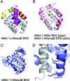
X-ray and NMR structures of the Mcl-1BLR:mNoxaB complex. (A) Ribbon diagram showing the mNoxaB BH3 peptide in purple and the BH1, BH2, and BH3 regions of hMcl-1 in blue, yellow, and red, respectively. (B) Overlay of hMcl-1BLR from the hMcl-1BLR:hBim BH3 complex (gray) and hMcl-1BLR:mNoxaB BH3 complex (pink). (C) Overlay of 20 minimum-energy NMR structures of the mMclBLR:mNoxaB BH3 complex. Mcl-1 is in blue, and the mNoxaB BH3 is in gray. (D) Close-up of the N terminus of the mNoxaB BH3 peptide from the NMR-derived structure illustrates that Glu-74 in the peptide engages Lys-215 of mMcl-1 (mMcl-1 numbering).
Fig. 4.
Fig. 4.
Mapping the region of the Noxa BH3 that induces Mcl-1 degradation. (A) BH3 domain chimeras of hBim and hNoxa. The signature sequence of four hydrophobic residues is indicated. The Bim BH3 sequence is underlined. (B) Human Noxa variants containing chimeric Noxa/Bim BH3 domains demonstrate that the C terminus of the BH3 is required for Noxa-induced Mcl-1 degradation.

References

    1. Adams JM. Genes Dev. 2003;17:2481–2495. - PubMed
    1. Muchmore SW, Sattler M, Liang H, Meadows RP, Harlan JE, Yoon HS, Nettesheim D, Chang BS, Thompson CB, Wong S, et al. Nature. 1996;381:335–341. - PubMed
    1. Petros AM, Medek A, Nettesheim DG, Kim DH, Yoon HS, Swift K, Matayoshi ED, Oltersdorf T, Fesik SW. Proc Natl Acad Sci USA. 2001;98:3012–3017. - PMC - PubMed
    1. Hinds MG, Lackmann M, Skea GL, Harrison PJ, Huang DCS, Day CL. EMBO J. 2003;22:1497–1507. - PMC - PubMed
    1. Day CL, Chen L, Richardson SJ, Harrison PJ, Huang DCS, Hinds MG. J Biol Chem. 2005;280:4738–4744. - PubMed

Publication types

MeSH terms