. 2007 May 11;282(19):14186-93.
doi: 10.1074/jbc.M700827200. Epub 2007 Mar 27.

Mitochondria are a major source of paraquat-induced reactive oxygen species production in the brain

Affiliations

Mitochondria are a major source of paraquat-induced reactive oxygen species production in the brain

Pablo R Castello et al. J Biol Chem. .

Abstract

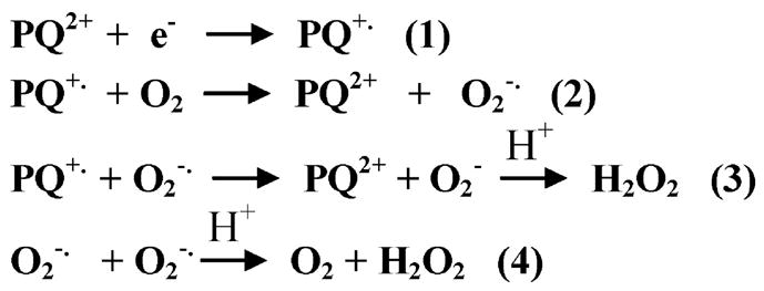
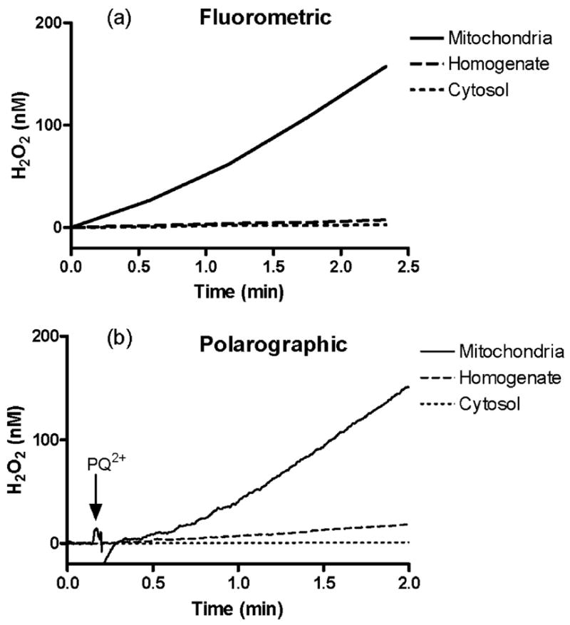
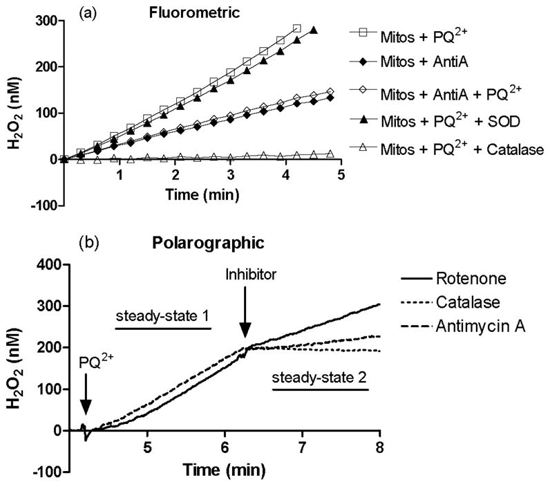
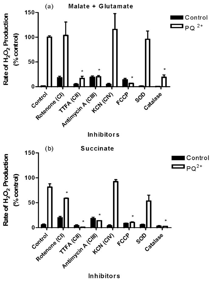
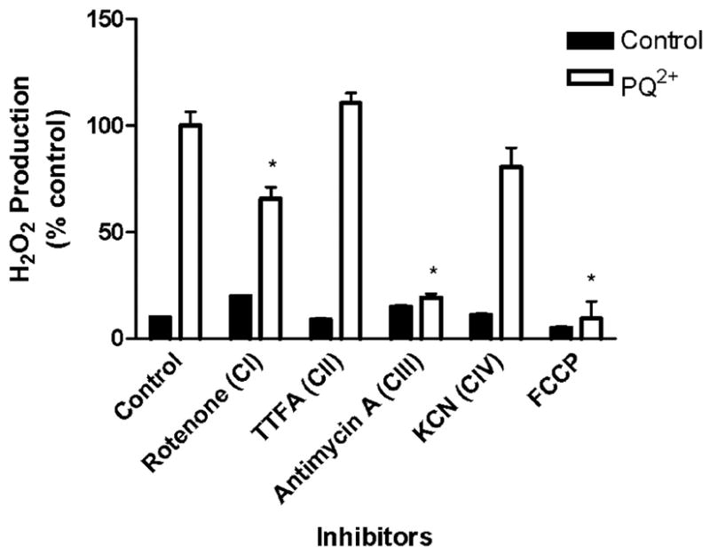
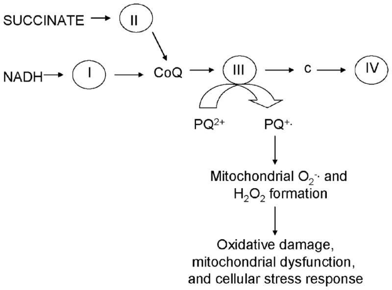
Paraquat (PQ(2+)) is a prototypic toxin known to exert injurious effects through oxidative stress and bears a structural similarity to the Parkinson disease toxicant, 1-methyl-4-pheynlpyridinium. The cellular sources of PQ(2+)-induced reactive oxygen species (ROS) production, specifically in neuronal tissue, remain to be identified. The goal of this study was to determine the involvement of brain mitochondria in PQ(2+)-induced ROS production. Highly purified rat brain mitochondria were obtained using a Percoll density gradient method. PQ(2+)-induced hydrogen peroxide (H(2)O(2)) production was measured by fluorometric and polarographic methods. The production of H(2)O(2) was evaluated in the presence of inhibitors and modulators of the mitochondrial respiratory chain. The results presented here suggest that in the rat brain, (a) mitochondria are a principal cellular site of PQ(2+)-induced H(2)O(2) production, (b) PQ(2+)-induced H(2)O(2) production requires the presence of respiratory substrates, (c) complex III of the electron transport chain is centrally involved in H(2)O(2) production by PQ(2+), and (d) the mechanism by which PQ(2+) generates H(2)O(2) depends on the mitochondrial inner transmembrane potential. These observations were further confirmed by measuring PQ(2+)-induced H(2)O(2) production in primary neuronal cells derived from the midbrain. These findings shed light on the mechanism through which mitochondria may contribute to ROS production by other environmental and endogenous redox cycling agents implicated in Parkinson's disease.


Figures

FIGURE 1
FIGURE 1
Chemical reactions proposed to participate in mechanisms of H2O2 production by PQ2+.
FIGURE 2
FIGURE 2. PQ2+-induced H2O2 production in cellular fractions from the brain
Fluorometric (a) and polarographic (b) assays were used to measure H2O2 in the presence of malate and glutmate following the addition of 250 μM PQ2+ to equal amounts of protein from each rat brain fraction: mitochondria (solid line), cytosol (dotted line), and homogenate (dashed line).
FIGURE 3
FIGURE 3. Inhibition of PQ2+-induced H2O2 production in stimulated mitochondria
Fluorometric (a) measurement of H2O2 production from isolated mitochondria in the presence of succinate was measured in the presence of PQ2+(250 μM, □), antimycin A (◆), antimycin A and PQ2+(◇), PQ2+ and SOD (▲), and PQ2+and catalase (△). Polarographic (b) measurement was used to confirm results. Following the addition of 250 μM PQ2+, H2O2 production in mitochondria was monitored with inhibitors added as indicated: rote-none (solid line), catalase (dotted line), and antimycin A (dashed line). Rates of H2O2 production before and after the addition of inhibitor (steady state 1 and 2, respectively) were compared with obtain the percentage of H2O2 production under each condition (see Fig. 5).
FIGURE 4
FIGURE 4. Effect of ETC inhibitors on PQ2+-induced H2O2 production in isolated mitochondria
Mitochondria were stimulated with respiration substrates (malate plus glutamate (a) or succinate (b)), and H2O2 production was measured in the absence (closed bars) or presence (open bars) of 250 μM PQ2+. Inhibitors were added as described under “Experimental Procedures.” H2O2 production in mitochondrial fraction stimulated with malate plus glutamate as respiration substrates in the presence of PQ2+ was considered 100%, and all values are expressed as a percentage of this (mean ±S.E., n =3). *, p <0.05 compared with PQ2+-treated control mitochondria (one-way analysis of variance).
FIGURE 5
FIGURE 5. Effect of ETC inhibitors on PQ2+-induced H2O2 production in primary midbrain cultures
H2O2 production was measured in midbrain cultures incubated in buffer containing PQ2+ and inhibitors where indicated using a fluorometric method as described under “Experimental Procedures.” H2O2 production in the presence of PQ2+ was considered 100%, and all values are expressed as a percentage of this (mean ± S.E., n = 3). *, p < 0.05 compared with PQ2+-treated control mitochondria (one-way analysis of variance).
FIGURE 6
FIGURE 6. Proposed involvement of ETC in PQ2+-dependent H2O2 generation in brain mitochondria
Electrons feed into the ETC at the level of complex I or complex II and are transferred to ubiquinone (CoQ), complex cytochrome c (c), and complex IV sequentially. Complex III has the ability transfer these electrons to PQ2+, thus forming the PQ radical, leading ROS formation and subsequent cell damage.

References

    1. Jenner P. Ann Neurol. 2003;53(Suppl 3):S26–S36. - PubMed
    1. Fahn S. Ann N Y Acad Sci. 2003;991:1–14. - PubMed
    1. Dauer W, Przedborski S. Neuron. 2003;39:889–909. - PubMed
    1. Bove J, Prou D, Perier C, Przedborski S. NeuroRx. 2005;2:484–494. - PMC - PubMed
    1. Bus JS, Gibson JE. Environ Health Perspect. 1984;55:37–46. - PMC - PubMed

Publication types

LinkOut - more resources