Review
. 2007;35(7):2295-301.
doi: 10.1093/nar/gkm104. Epub 2007 Mar 27.

Ribosomal RNA guanine-(N2)-methyltransferases and their targets

Affiliations
Review

Ribosomal RNA guanine-(N2)-methyltransferases and their targets

Petr V Sergiev et al. Nucleic Acids Res. 2007.

Abstract

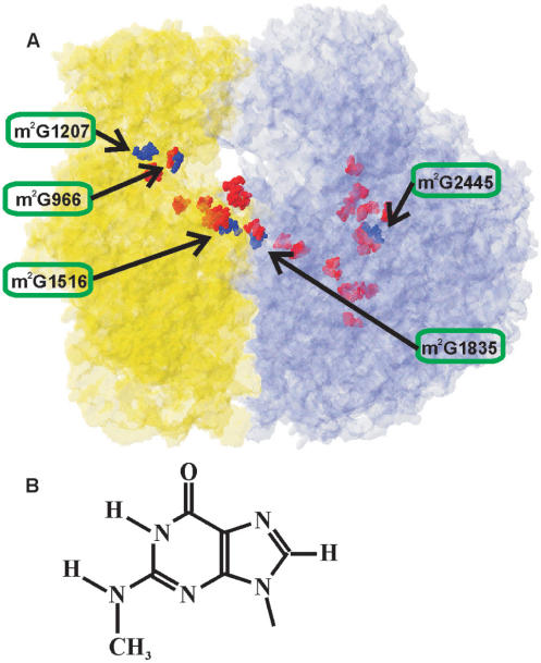
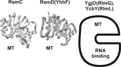
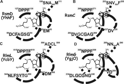
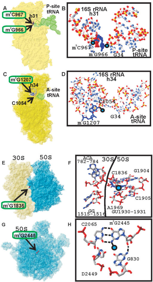
Five nearly universal methylated guanine-(N2) residues are present in bacterial rRNA in the ribosome. To date four out of five ribosomal RNA guanine-(N2)-methyltransferases are described. RsmC(YjjT) methylates G1207 of the 16S rRNA. RlmG(YgjO) and RlmL(YcbY) are responsible for the 23S rRNA m(2)G1835 and m(2)G2445 formation, correspondingly. RsmD(YhhF) is necessary for methylation of G966 residue of 16S rRNA. Structure of Escherichia coli RsmD(YhhF) methyltransferase and the structure of the Methanococcus jannaschii RsmC ortholog were determined. All ribosomal guanine-(N2)-methyltransferases have similar AdoMet-binding sites. In relation to the ribosomal substrate recognition, two enzymes that recognize assembled subunits are relatively small single domain proteins and two enzymes that recognize naked rRNA are larger proteins containing separate methyltransferase- and RNA-binding domains. The model for recognition of specific target nucleotide is proposed. The hypothetical role of the m(2)G residues in rRNA is discussed.


Figures

Figure 1.
Figure 1.
(A) Modified nucleotides distribution in the structure of the E. coli ribosome (27). Small subunit is shown in yellow; large subunit is shown in blue. N2-methylguanosine residues are shown as dark blue Van-der-Vaals spheres. The residue number of m2G nucleotides in 16S or 23S rRNA is depicted and the location of particular m2G residue is indicated by arrow. Other modified nucleotides are shown as red Van-der-Vaals spheres. (B) The structure of N2-methylguanine.
Figure 2.
Figure 2.
The structure of ribosomal guanine-(N2)-methyltransferases. The structure of Mj0882 protein (12), which is likely to represent an ortholog of E. coli RsmC is marked as ‘RsmC’. The structure of E. coli RsmD(YhhF) protein is marked accordingly. Both protein structures are also marked ‘MT’ to indicate that they consist of single methyltransferase domain. For RlmG(YgjO) and RlmL(YcbY) the scheme is shown, where the protein is divided into N-terminal RNA-binding domain and C-terminal methyltransferase (MT) domain, based on conserved domain analysis (38).
Figure 3.
Figure 3.
The scheme of AdoMet-binding site of the ribosomal guanine-(N2)-methyltransferases. (A) RsmD(YhhF) active site, (B) RsmC(YjjT) active site, (C) RlmL(YcbY) active site and (D) RlmG(YgjO) active site. AdoMet structure is shown in the center of each scheme. It is surrounded by protein sequences, proximal to the putative AdoMet-binding site. Conserved amino acids are shaded gray. Probable hydrogen bonds are indicated by dotted lines. Hypothetical stacking interactions of the adenine base are shown by dashed lines. Open circles indicate the location of amino acid side chain below the adenine plane; closed circles indicate that amino acid is stacked above the adenine plane.
Figure 4.
Figure 4.
Fitting of RsmD(YhhF) structure (dark gray) to the P-site of 3D structure of the small ribosomal subunit (light gray). T. thermophilus 30S subunit in open conformation (15), was used for the fitting. The head of the small subunit was moved upward by 4 Å to avoid steric clashes. This movement is highly probable in vivo given high conformational mobility of the small subunit head.
Figure 5.
Figure 5.
Location of the m2G966, m2G1207 residues of the 16S rRNA and m2G1835, m2G2445 residues of the 23S rRNA in the ribosomal structure and their interactions. (A) Position of the 16S rRNA residue m2G966 relative to the P-site-bound tRNA (17). The 30S subunit is shown in yellow. P-site-bound tRNA is shown in green. Helix 31 is indicated as a gray tubing. Modified nucleotides m2G966 and m2G967 are shown as a blue and red Van-der-Vaals spheres accordingly and labeled. (B) Closer view of the position of the 16S rRNA residue m2G966 relative to the P-site-bound tRNA. 16S rRNA helix 31 is shown on the left, P-site-bound tRNA anticodon is shown on the right. (C) Position of the 16S rRNA residue m2G1207 relative to the A-site-bound tRNA anticodon (23). The 30S subunit is shown in yellow. A-site-bound tRNA anticodon is shown in green. Helix 34 is indicated as a gray tubing. Modified nucleotide m2G1207 is shown as red Van-der-Vaals spheres and labeled. Nucleotide C1054 making a direct contact with A-site tRNA anticodon is shown as wireframe and labeled. (D) Closer view of the position of the 16S rRNA residue m2G1207 relative to the A-site-bound tRNA. 16S rRNA helix 34 is shown on the left, A-site-bound tRNA anticodon is shown on the right. (E) Position of the 23S rRNA residue m2G1835 in the E. coli ribosome (27). Position of the m2G1835 residue relative to the ribosomal subunits. The 30S subunit is shown in yellow; the 50S subunit is shown in blue. Modified nucleotide m2G1835 is shown as red Van-der-Vaals spheres and labeled. (F) Details of structural environment of the m2G1835 in the ribosome. Position of the methyl group is marked by blue circle. Surrounding nucleotides are shown as wireframe and labeled. 16S rRNA nucleotides are on the left side, 23S rRNA nucleotides are on the right side. Border between the subunits is indicated by line. (G) Position of the 23S rRNA residue m2G2445 in the E. coli 50S subunit (27). Position of the m2G2445 residue relative to the large ribosomal subunit, shown in blue and labeled. The orientation of the 50S subunit is as viewed from the 30S subunit. (H) Details of structural environment of the m2G2445 in the 50S subunit. Position of the methyl group is marked by blue circle. Surrounding nucleotides are shown as wireframe and labeled.

Similar articles

Cited by

References

    1. Andersen NM, Douthwaite S. YebU is a m5C methyltransferase specific for 16S rRNA nucleotide 1407. J. Mol. Biol. 2006;359:777–786. - PubMed
    1. Mueller F, Brimacombe R. A new model for the three-dimensional folding of Escherichia coli 16S Ribosomal RNA. I. Fitting the RNA to a 3D electron microscopic map at 20 Å. J. Mol. Biol. 1997;271:524–544. - PubMed
    1. Cundliffe E. Ribosomal modification and resistance in antibiotic-producing organisms. Biochem. Soc. Symp. 1987;53:1–8. - PubMed
    1. Maravic G. Macrolide resistance based on the Erm-mediated rRNA methylation. Curr. Drug Targets Infect. Disord. 2004;4:193–202. - PubMed
    1. Del Campo M, Kaya Y, Ofengand J. Identification and site of action of the remaining four putative pseudouridine synthases in Escherichia coli. RNA. 2001;7:1603–1615. - PMC - PubMed

Publication types

MeSH terms