

Age-related differences in the normal human cornea: a laser scanning in vivo confocal microscopy study
- PMID: 17389741
- PMCID: PMC1954900
- DOI: 10.1136/bjo.2006.112656
Age-related differences in the normal human cornea: a laser scanning in vivo confocal microscopy study
Abstract
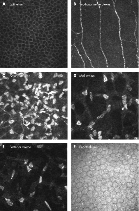
Aims: To quantify and establish baseline normative data for age-related differences in cellular and innervation density in the normal, healthy, human cornea using laser scanning in vivo confocal microscopy.
Methods: Cross-sectional study of 85 normal subjects assessed via corneal topography and laser scanning in vivo confocal microscopy.
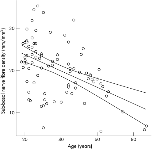
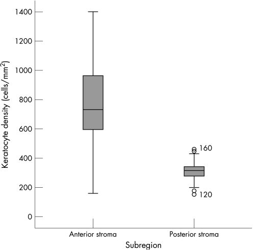
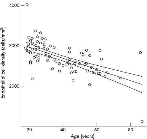
Results: Mean age was 38+/-16 years (range 18-87 years) and 60% of subjects were female. Anterior keratocyte density declined by 0.9% per year (r = -0.423, p<0.001), posterior keratocyte density declined by 0.3% per year (r = -0.250, p = 0.021) and endothelial cell density declined by 0.5% per year (r = -0.615, p<0.001). Sub-basal nerve fibre density declined by 0.9% per year (r = -0.423, p<0.001). No association was observed between age and basal epithelial cell density, or between age and central corneal thickness, corneal astigmatism or horizontal corneal diameter (p>0.05). No association was observed between subject gender and corneal cell or innervation density.
Conclusions: Using laser scanning in vivo confocal microscopy this study highlights a significant, and relatively linear, reduction in keratocyte and endothelial cell density with increasing subject age. Interestingly, corneal sub-basal nerve fibre density also significantly decreases with increasing age. In vivo laser scanning confocal microscopy provides a safe, non-invasive method for the establishment of normative data and assessment of alterations in human corneal microstructure following surgery or disease processes.
Conflict of interest statement
Competing interests: None declared
References
-
- Chatterjee A, Shah S S, Doyle S J. Effect of age on final refractive outcome for 2342 patients following photorefractive keratectomy. Invest Ophthalmol Vis Sci 199637S57
-
- Marre M. On the age dependence of the healing of corneal epithelium defects (In German). Albrecht Von Graefes Arch Klin Exp Ophthalmol 1967173250–255. - PubMed
MeSH terms
LinkOut - more resources
Full Text Sources
Medical