. 2007 Mar 28;2(3):e318.
doi: 10.1371/journal.pone.0000318.

Cohesin is dispensable for centromere cohesion in human cells

Affiliations

Cohesin is dispensable for centromere cohesion in human cells

Laura A Díaz-Martínez et al. PLoS One. .

Abstract

Background: Proper regulation of the cohesion at the centromeres of human chromosomes is essential for accurate genome transmission. Exactly how cohesion is maintained and is then dissolved in anaphase is not understood.

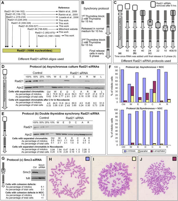
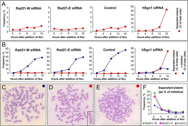
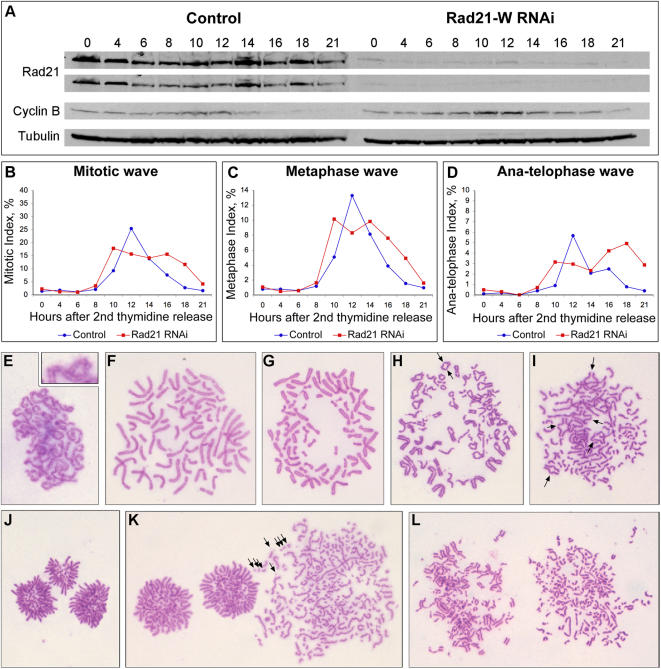
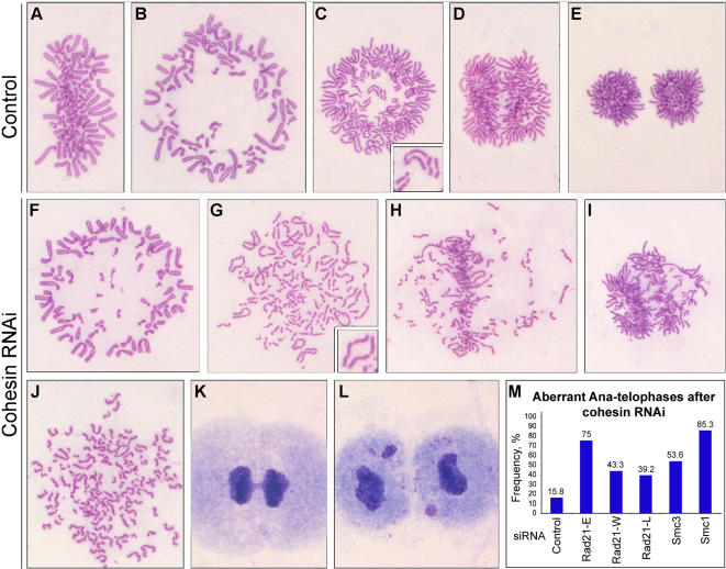
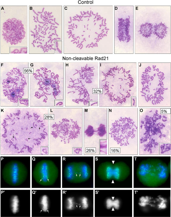
Principal findings: We have investigated the role of the cohesin complex at centromeres in human cells both by depleting cohesin subunits using RNA interference and also by expressing a non-cleavable version of the Rad21 cohesin protein. Rad21 depletion results in aberrant anaphase, during which the sister chromatids separate and segregate in an asynchronous fashion. However, centromere cohesion was maintained before anaphase in Rad21-depleted cells, and the primary constrictions at centromeres were indistinguishable from those in control cells. Expression of non-cleavable Rad21 (NC-Rad21), in which the sites normally cleaved by separase are mutated, resulted in delayed sister chromatid resolution in prophase and prometaphase, and a blockage of chromosome arm separation in anaphase, but did not impede centromere separation.

Conclusions: These data indicate that cohesin complexes are dispensable for sister cohesion in early mitosis, yet play an important part in the fidelity of sister separation and segregation during anaphase. Cleavage at the separase-sensitive sites of Rad21 is important for arm separation, but not for centromere separation.


Conflict of interest statement

Competing Interests: The authors have declared that no competing interests exist.

Figures

Figure 1
Figure 1. Rad21 depletion in HeLa cells.
(A) Cartoon showing locations in the Rad21 mRNA sequence that were targeted by 9 different siRNA oligos used in this study. (B) Protocol used for achieving early S-phase synchrony of HeLa cells. (C) Summary of 8 different protocols used to deplete Rad21. Open rectangle indicates the timing of siRNA transfection relative to the synchrony protocol (which was done as described in B) or in asynchronous culture (leftmost arrow). Protocol-A, -B and -C are indicated as (a, b, c). For the protocols involving synchrony, the numbers at the bottom of each protocol indicate the total time from the siRNA transfection until the cells were released from the final early S-phase arrest, at which time protein extracts were prepared for biochemical analysis. In parallel, samples were fixed with Carnoy's for cytological analysis 11 hours after the release (in half of the samples, nocodazole was present during the last 2 hours before Carnoy's fixation). For the asynchronous protocol, protein samples and Carnoy's fixed samples were prepared 48 hours after siRNA transfection (in half of the samples, nocodazole was present for 2 additional hours and thus the cells were fixed with Carnoy's 50 hours after siRNA transfection). (D,E) Western blots showing degrees of Rad21 depletion achieved using Protocol-A and Protocol-B. Apc2 is a loading control. Left four lanes are dilution series of control samples. Letters above the other lanes denote the siRNA oligos used and correspond to those depicted in A. (F, H–J) Cytological analysis corresponding to the biochemical analysis shown in D and E. Samples were scored to determine the frequencies of mitotic cells that had cohered sisters (H), resolved primary constrictions (I) or fully separated sister chromatids (J). (Note that the cells shown are not arrested in nocodazole, and therefore serve as examples only. Examples with nocodazole are shown in Fig. 2.) The data tabulated in D and E indicate the frequencies of sister separation (combining the categories depicted in I and J) and the histogram plots (F) are colored corresponding to the categories depicted in H–J. (G) Depletion of Smc3 in HeLa cells (Protocol-C). Western blot shows Smc3 level at the time of release from S-phase synchrony. Tub = alpha-tubulin loading control. Cells with separated sisters chromatids (in tabulated form) were quantified from chromosome preparations fixed with Carnoy's and stained with Giemsa.
Figure 2
Figure 2. Rad21 depleted cells do not separate their sister chromatids in the presence of nocodazole.
(A–E) HeLa cells synchronized in early S-phase and depleted of Rad21 (or mock depleted) with the indicated siRNA oligos as described in Figure 1, Protocol-B. Nocodazole was added after release from the S-phase synchrony and cells prepared for cytological analysis at the indicated time points. Cells with separated or partly separated sister chromatids were scored versus cells with cohered sisters, as depicted in C–E. For comparison, Sgo1-depleted cells were examined under the same conditions. Sgo1 depletion (∼80% depletion) was slightly less efficient that Rad21 depletion (data not shown). Note that the data in panel A are reproduced in panel B but with a different Y-axis scale. (F) HeLa cells were depleted of Rad21 (or mock depleted) with the indicated siRNA oligos as described in Figure 1, Protocol-A. Nocodazole was added 48 hours after siRNA transfection and cells prepared for cytological analysis at the indicated time points, then scored as in A–E (% separated sisters combines the categories shown in D+E).
Figure 3
Figure 3. Time course of synchronous Rad21-depleted HeLa cells (Rad21-W siRNA).
HeLa cells were synchronized by double thymidine block and transfected (or mock transfected) with Rad21-W siRNA according to Protocol-C (see Figure 1). After release from the early S-phase synchrony, samples were taken for biochemical (A) and cytological (B–L) analysis over the next 21 hours. (A) Western blot showing level of Rad21 depletion and mitotic status of the cells based on cyclin B levels (Tubulin = loading control). (B–D) Samples for cytology were fixed and stained (see Material and Methods) and mitotic categories scored on at least 1000 cells per sample and time point. (E–F) Cytological features of cells transfected with Rad21-W siRNA: (E) Normal sister cohesion upon nuclear envelope breakdown (observed in 100% of the prophase and prometaphase cells); (F) Normal sister cohesion in early prometaphase; (G) Normal metaphase plate formation; (H–I) Aberrant anaphases – centromere regions separating before arms (arrows) and some chromosomes segregating to the poles before other chromosomes have separated their sisters. (J) Abnormal telophase – chromosomes have segregated (unevenly) to three cell poles; (K) Chromosome breaks in a cell with separated sister chromatids (presumably a post-anaphase cell); (L) Chromosome breaks in metaphase or early anaphase cells.
Figure 4
Figure 4. Anaphase and telophase abnormalities in Rad21-depleted HeLa cells.
(A–L) Cells fixed with Carnoy's and stained with Giemsa. (A–E) Control series showing the cytology of control-treated cells at different mitotic stages: (A,B) Metaphases - side and polar views; (C) Early anaphases – polar and side views; (E) Telophase. (F–L) Examples of cells treated with Rad21-specific siRNA: (F) Metaphase – polar view; (G–I) Aberrant anaphase – asynchronous anaphase, defined as described in Fig. 3; (J) Apolar telophase – separated sisters scattered in the cytoplasm (cell may be arrested in mitosis based on the level of chromosome condensation); (K–L) Telophases with chromosome bridges and lagging chromosomes; (M) Quantification of abnormal anaphase and telophase cells in control-treated and cohesin-depleted cells. At least 1000 cells were scored per sample.
Figure 5
Figure 5. Cytological analysis of HeLa cells expressing non-cleavable (NC) Rad21.
Control HeLa cells in which wild type MYC-Rad21 expression had been induced (A–E) were compared with HeLa in which NC-Rad21 (NC-MYC-Rad21) expression had been induced 72 hours prior to fixation and staining with Giemsa (F–O) or immuno-staining with CREST serum (P–T). Controls: (A,B) Normal cohesion and sister resolution in late prophase and early prometaphase; (C) Metaphase – polar view; (D) Anaphase -side view; (E) Telophase. NC-Rad21: (F–G) Delayed nucleolus disassembly in early prometaphase and delayed sister chromatid resolution (56% of prometaphases had the resolution defect); (H–I) Late prometaphase and metaphase cells with unresolved sisters (32%); (J) Metaphase – polar view; (K–L) Aberrant anaphases – centromere regions separating before arms (arrows) and some chromosomes segregating to the poles before other chromosomes have separated their sisters (28% were abnormal based on these criteria); (M) Cut telophase (26% of the telophases had this phenotype); (N) Apolar telophase (16% of the telophases had this phenotype); (O) Partially resolved diplochromosomes (5% of mitotics), in agreement with the described separase RNAi phenotype ; (P–T) DNA (DAPI; blue) and CREST (green) staining of kinetochores after NC-Rad21 induction. (P′–T′) DAPI channel only. (P) Metaphase; (Q) Early anaphase – one pair of kinetochores appears to be segregating prematurely; (R) Laggard kinetochores in anaphase; (S) Cut telophase - kinetochores have segregated to the poles despite deficient karyokinesis; (T) Bridged and laggard chromosomes in telophase.

Similar articles

Cited by

References

    1. Nasmyth K. How might cohesin hold sister chromatids together? Philos Trans R Soc Lond B Biol Sci. 2005;360:483–496. - PMC - PubMed
    1. Huang CE, Milutinovich M, Koshland D. Rings, bracelet or snaps: fashionable alternatives for Smc complexes. Philos Trans R Soc Lond B Biol Sci. 2005;360:537–542. - PMC - PubMed
    1. Uhlmann F, Nasmyth K. Cohesion between sister chromatids must be established during DNA replication. Curr Biol. 1998;8:1095–1101. - PubMed
    1. Losada A, Yokochi T, Kobayashi R, Hirano T. Identification and characterization of SA/Scc3p subunits in the Xenopus and human cohesin complexes. J Cell Biol. 2000;150:405–416. - PMC - PubMed
    1. Sumara I, Vorlaufer E, Gieffers C, Peters BH, Peters JM. Characterization of vertebrate cohesin complexes and their regulation in prophase. J Cell Biol. 2000;151:749–762. - PMC - PubMed

Publication types

MeSH terms