. 2007 May 7:1148:1-14.
doi: 10.1016/j.brainres.2007.02.069. Epub 2007 Mar 3.

Mutational analysis of aspartoacylase: implications for Canavan disease

Affiliations

Mutational analysis of aspartoacylase: implications for Canavan disease

Jeremy R Hershfield et al. Brain Res. .

Abstract

Mutations that result in near undetectable activity of aspartoacylase, which catalyzes the deacetylation of N-acetyl-l-aspartate, correlate with Canavan Disease, a neurodegenerative disorder usually fatal during childhood. The underlying biochemical mechanisms of how these mutations ablate activity are poorly understood. Therefore, we developed and tested a three-dimensional homology model of aspartoacylase based on zinc dependent carboxypeptidase A. Mutations of the putative zinc-binding residues (H21G, E24D/G, and H116G), the general proton donor (E178A), and mutants designed to switch the order of the zinc-binding residues (H21E/E24H and E24H/H116E) yielded wild-type aspartoacylase protein levels and undetectable ASPA activity. Mutations that affect substrate carboxyl binding (R71N) and transition state stabilization (R63N) also yielded wild-type aspartoacylase protein levels and undetectable aspartoacylase activity. Alanine substitutions of Cys124 and Cys152, residues indicated by homology modeling to be in close proximity and in the proper orientation for disulfide bonding, yielded reduced ASPA protein and activity levels. Finally, expression of several previously tested (E24G, D68A, C152W, E214X, D249V, E285A, and A305E) and untested (H21P, A57T, I143T, P183H, M195R, K213E/G274R, G274R, and F295S) Canavan Disease mutations resulted in undetectable enzyme activity, and only E285A and P183H showed wild-type aspartoacylase protein levels. These results show that aspartoacylase is a member of the caboxypeptidase A family and offer novel explanations for most loss-of-function aspartoacylase mutations associated with Canavan Disease.


Figures

Fig. 1
Fig. 1. Sequence alignment between ASPA and bovine ZnCPA (PDB structure 8CPA)
The numbers indicated in parenthesis for the ASPA gene begin with the glutamine residue that aligns with proline 60 of ZnCPA. The conserved residues that are possibly involved in a zinc-dependent catalytic mechanism for ASPA are shown white in blue boxes with the putative zinc ligands highlighted by asterisks. The line above the alignment indicates the secondary structure (H for α-helix, E for extended, and T for turn) as reported with the 8CPA crystal structure (http://www.rcsb.org). Dashes denote amino acids that do not align with 8CPA. Residues predicted to be involved in catalysis are numbered below the alignment.
Fig. 2
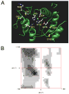
Fig. 2. Partial 3D homology modeling of ASPA based on the crystal structure of bovine ZnCPA
A, The homology model for ASPA. Some of the loops are shown for clarity. The residues of interest in this report are depicted as ball-and-stick. A single zinc atom has been placed in the active site as a cyan sphere. B, Ramachandran Plot of the backbone torsion angles (phi and psi) for the homology model.
Fig. 3
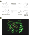
Fig. 3. Comparison of reactions and active sites of ASPA and bovine ZnCPA
A, Chemical reactions catalyzed by ASPA and ZnCPA. The arrows point to hydrolyzed peptide bonds. B, Superposition of critical active site residues between ASPA and ZnCPA. The ASPA residues are depicted in atom color-coded ball-and-stick. The bovine ZnCPA residues are overlayed in thick yellow lines. Labeling corresponds to ZnCPA numbering.
Fig. 4
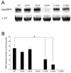
Fig. 4. Mutational analysis of ASPA residues that align with those involved in bovine ZnCPA’s catalytic mechanism
Whole cell extracts prepared from COS-7 cells transiently transfected with the indicated constructs were analyzed by immunoblotting and radiometric ASPA assay (see Materials and Methods). The enzyme activities are presented as mean +/− SEM for triplicate transfections. A, Immunoblotting using pepASPA and α-βT. B, Each putative catalytic mutation resulted in undetectable ASPA activity relative to WT. C, Changing the order of the putative Zn-binding ligands results in undetectable ASPA activity.
Fig. 5
Fig. 5. Purification of human ASPA from E. coli
The lysate and the indicated eluates of bacteria transformed with the indicated constructs were analyzed by SDS-PAGE and Coomassie staining (see Materials and Methods). Recombinant human ASPA fused to N-terminal thioredoxin and C-terminal V5 and (6x)-histidine tags was ~52 kDa.
Fig. 6
Fig. 6. Cysteine mutational analysis indicates a possible disulfide bond in hASPA
Whole cell extracts prepared from COS-7 cells transiently transfected with the indicated constructs were analyzed by immunoblotting and radiometric ASPA assay (see Materials and Methods). The enzyme activities are presented as mean +/− SEM for six transfections. A, Immunoblotting using pepASPA and α-βT. B, Expression of C61A and C61S resulted in WT ASPA activity, expression of C124A and C152A resulted in statistically significantly reduced activity levels relative to WT (*; p < 0.05), and expression of C61W and C152W resulted in undetectable activity.
Fig. 7
Fig. 7. CD mutations give insight into hASPA structure
Whole cell extracts prepared from COS-7 cells transiently transfected with the indicated constructs were analyzed by immunoblotting and radiometric ASPA assay (see Materials and Methods). The enzyme activities are presented as mean +/− SEM for triplicate transfections. ND, No DNA mock transfection.. A, Analysis of select previously in vitro tested CD mutations. Top panel: RT-PCR confirmed that each mutant construct was expressed comparably to WT hASPA. Middle panel: Immunoblotting using a polyclonal peptide antibody (pepASPA) and β-tubulin loading control (α-βT). The ASPA band is 38 kDa and the beta-tubulin band is 50 kDa. Bottom panel: In vitro expression of each CD mutant resulted in undetectable ASPA activity compared to WT. B, Analysis of select previously in vitro untested CD mutations. Top panel: RT-PCR confirmed that each construct was expressed comparably to WT. Middle panel: Immunoblotting using pepASPA and α-βT. Bottom panel: Each mutant resulted in undetectable activity, with the following exceptions: WT activity for K213E and ~5% residual activity for G274R. C, Analysis of a case report of clinically mild CD. Top panel: Immunoblotting using pepASPA and α-βT. K213E/G274R is a double mutant expressed from a single plasmid (see Materials and Methods). Bottom panel: Expression of both G274R and the K213E/G274R double mutant in this set of triplicate transfections resulted in undetectable ASPA activity compared to WT and K213E.
Fig. 8
Fig. 8. Proposed catalytic mechanism for NAA hydrolysis by ASPA
A, NAA substate binding. B, Nucleophilic attack. C, Transition state intermediate. D, Acetate and aspartate product formation.

References

    1. Auld DS. Zinc coordination sphere in biochemical zinc sites. Biometals. 2001;14:271–313. - PubMed
    1. Benuck M, D’Adamo AF., Jr Acetyl transport mechanisms. Metabolism of N-acetyl-L-aspartic acid in the non-nervous tissues of the rat. Biochim Biophys Acta. 1968;152:611–618. - PubMed
    1. Berman HM, Westbrook J, Feng Z, Gilliland G, Bhat TN, Weissig H, Shindyalov IN, Bourne PE. The Protein Data Bank. Nucleic Acids Res. 2000;28:235–242. - PMC - PubMed
    1. Bitto E, Bingman CA, Wesenberg GE, McCoy JG, Phillips GN., Jr Structure of aspartoacylase, the brain enzyme impaired in Canavan disease. Proc Natl Acad Sci U S A. 2007;104:456–461. - PMC - PubMed
    1. Bukrinsky JT, Bjerrum MJ, Kadziola A. Native carboxypeptidase A in a new crystal environment reveals a different conformation of the important tyrosine 248. Biochemistry. 1998;37:16555–16564. - PubMed

Publication types

MeSH terms

LinkOut - more resources