. 2007 Apr;170(4):1415-27.
doi: 10.2353/ajpath.2007.060728.

Autoantibodies to vimentin cause accelerated rejection of cardiac allografts

Affiliations

Autoantibodies to vimentin cause accelerated rejection of cardiac allografts

Balakrishnan Mahesh et al. Am J Pathol. 2007 Apr.

Abstract

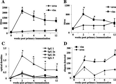
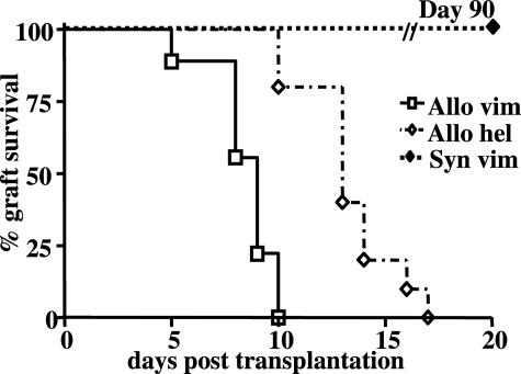
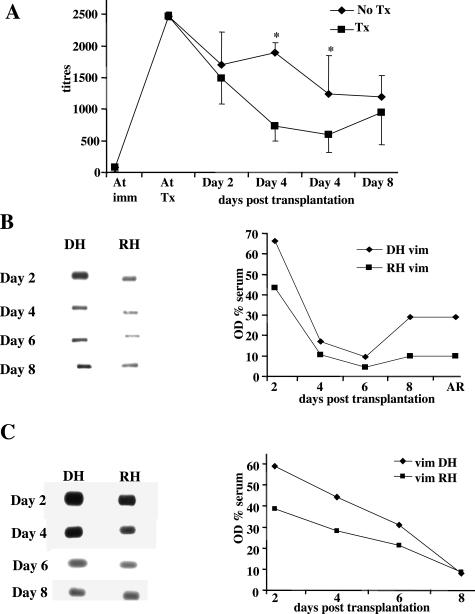
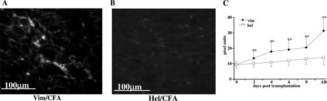
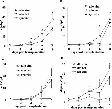
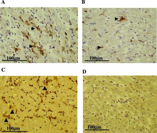
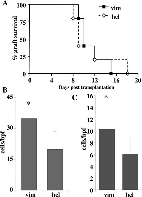
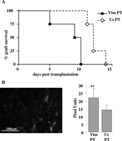
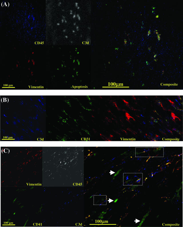
Autoimmune responses to vimentin occur after solid organ transplantation, but their pathogenic effects are unclear. The aim of these studies was to investigate the effects of vimentin preimmunization on allogeneic and isografted hearts in a murine transplant model. Immunization of C57BL/6 mice with murine vimentin in complete Freund's adjuvant resulted in anti-vimentin antibodies and vimentin-reactive Th-1 cells. Transplantation of 129/sv hearts into vimentin-immunized C57BL/6 recipients resulted in accelerated rejection (8.4 +/- 1.5 days; n = 18), compared with hen egg lysozyme-immunized C57BL/6 (13.3 +/- 2.2 days; n = 10; P < 0.0001, log-rank test). In contrast, isografts continued to beat beyond 90 days. Immunohistochemical analysis of allografts from vimentin/complete Freund's adjuvant mice demonstrated increased numbers of T cells and enhanced microvascular deposition of C3d, CD41, and P-selectin compared with controls. Antibodies were necessary for accelerated rejection, shown by the fact that vimentin-immunized B-cell-deficient IgH6 mice did not show accelerated rejection of 129/sv allografts, but rejection was restored by adoptive transfer of serum containing anti-vimentin antibodies. Eluates from donor hearts placed in vimentin/complete Freund's adjuvant recipients contained anti-vimentin antibodies, shown by Western blotting. Confocal imaging of rejected hearts de-monstrated presence of vimentin and C3d on apoptosed leukocytes, endothelial cells, and platelet/leukocyte conjugates. These results demonstrate that autoantibodies to vimentin, in conjunction with the alloimmune response, have a pathogenic role in allograft rejection.


Figures

Figure 1
Figure 1
Antibody and cytokine production in vimentin-immunized mice. C57BL/6 mice were immunized with 400 μg of vimentin emulsified in CFA followed by a booster of 400 μg of vimentin alone 1 week later. Controls received 100 μl of 6 mol/L urea (vehicle) in CFA. ELISA was used to determine titers of IgG (A) and IgM (B) AVA at 2 to 12 weeks after the first injection. IgG subclasses of AVA were determined in C. Points represent means and standard deviations of four mice per group. *P < 0.05 for differences between vimentin-immunized mice and controls in A and B and for differences between IgG2b titers and other subclasses in C. D: Cytokine production by splenocytes from vimentin-immunized mice assessed by ELISPOT. Spleens from mice immunized with vim/CFA (vim) or urea/CFA (urea) were removed at time 0 and 2 to 12 weeks after the first injection, and splenocytes were prepared and cultured with murine vimentin to determine production of IFN-γ (see Materials and Methods). In brief, 106 splenocytes were cultured with vimentin, urea, or concanalavin-A (not shown) in vitro, in wells coated with mAb to IFN-γ (D). After 48 hours, the cells were lysed with distilled water and ELISPOTs developed using antibodies to appropriate biotinylated mAb. Points represent means and standard deviations of four mice per group. *P < 0.05 for differences between vimentin- or urea-immunized mice.
Figure 2
Figure 2
Accelerated rejection of cardiac allografts in vimentin-immunized mice. Kaplan-Meier survival graft shows accelerated rejection of 129/sv allografts placed in vim/CFA (Allo vim, n = 18) recipients compared with allografts placed in hel/CFA-immunized (Allo hel, n = 10) recipients (P < 0.0001, log-rank test). Isografts placed in vimentin-immunized C57BL/6 recipients (Syn vim) continued to beat at day 90. Vim/CFA mice were immunized with 400 μg of vimentin in 100 μl of CFA followed by a booster of 400 μg of vimentin alone without adjuvant. The hel/CFA controls received 200 μg of hel in 100 μl of CFA, followed by a booster of 200 μg of hel alone at 1 week. Recipients were transplanted 1 week after administration of booster antigen.
Figure 3
Figure 3
Levels of AVA in serum, transplanted hearts, and own hearts of vim/CFA-immunized mice. Time course of IgG AVA in serum of C57BL/6 mice immunized with vim/CFA and either receiving a 129/sv cardiac allograft (Tx) or not transplanted (No Tx), mean ± SD from five sera/group (A) (*P < 0.05). Western blot showing the presence of AVA in allografted 129/sv hearts (DH, B) or isografted C57BL/6 hearts (DH, C) placed in C57BL/6 mice immunized with vim/CFA (vim), and recipients’ own hearts (RH), removed at days 2, 4, 6, 8, and time of rejection. The OD % serum on the y axis represents the OD reading of the eluate from pooled hearts (either DH or RH) as a percentage of OD reading from pooled serum taken at the same time as hearts.
Figure 4
Figure 4
Deposition of C3d in cardiac allografts. Photomicrograph of frozen section of 129/sv cardiac allograft placed in vim/CFA- (A) or hel/CFA- (B) immunized recipients taken at time of rejection, stained with FITC-conjugated rabbit anti-human C3d. Quantification of C3d deposits at days 2 to 8 and time of acute rejection (AR) revealed more C3d deposition in allografts placed in vimentin-immunized C57BL/6 recipients compared with allografts placed in hel-immunized recipients (C). Quantification is expressed as pixel units on the y axis. **P < 0.05 compared with the preceding time point and compared with hel/CFA recipient.
Figure 5
Figure 5
Quantification of T cells and CD41 platelets. Quantification of CD3+ (A), CD4+ (B), and CD8+ (C) T cells and CD41+ platelets (D) infiltrating 129/sv allografts in C57BL/6 recipients immunized with vim/CFA (allo vim), hel/CFA (allo hel), or C57BL/6 isografts placed in vim/CFA (syn vim)-immunized mice at various times after transplantation. Results expressed as cells/hpf, mean ± SD (n = 4 mice/group). *P < 0.05 for differences between vim/CFA and hel/CFA controls.
Figure 6
Figure 6
Demonstration of endothelial cell activation and microvascular damage in cardiac allografts from vim/CFA recipients. Photomicrographs of frozen sections of cardiac allografts from vim/CFA (A and C) or hel/CFA (B and D) recipients stained with mAb to CD41 (A and B) or CD62P (P-selectin, C and D) 6 days after transplantation. Hearts from vim/CFA recipients (C) show extensive microvascular expression of P-selectin compared with hearts from hel/CFA recipients (D). Hearts from mice at day 4 show similar results to day 6 (not shown).
Figure 7
Figure 7
Rejection of allografts in vim/CFA IgH6 mice. Kaplan-Meier graft survival of 129/sv allografts placed in IgH6 immunized with vim/CFA (vim) or hel/CFA (hel) (A) demonstrates no difference in survival. Quantification of CD3 (B) and CD8 (C) T cells in cardiac allografts from immunized (vim) and hel mice (hel) showed increased CD3 and CD8 T cells in hearts of vim/CFA recipients at time of rejection. Cells expressed as numbers/hpf, mean ±SD (n = 4/group). *P < 0.05 for comparison of vim-immunized and hel-immunized groups.
Figure 8
Figure 8
Passive transfer (PT) of immunized serum restores accelerated rejection. Kaplan-Meier survival curve of 129/sv allografts placed in IgH6 mice, which received serum from immunized (Vim PT) or unimmunized rabbits (Us PT) (A). Quantification of C3d in hearts at times of rejection is shown in the histogram (B), and the photomicrograph shows immunocytochemical localization of C3d in frozen section of heart placed in vim/CFA recipient at the time of rejection. **P < 0.05 for differences between Vim PT and Us PT.
Figure 9
Figure 9
Confocal laser scanning microscopy of cryostat sections of allografted heart from vimentin-immunized recipient at time of rejection. Antibody combinations are described in Materials and Methods. In A, sections stained for leukocytes (CD45-APC), C3d (Cascade Blue, pseudostained for white to represent the C3d signal), vimentin (Alexa 594), and apoptosis (terminal deoxynucleotidyl transferase dUTP nick-end labeling-FITC). The composite shows co-localization of vimentin and C3d expression on apoptosing infiltrating recipient leukocytes. In B, sections were stained for C3d (Cascade Blue), endothelial cells, CD31 (Alexa 546), and vimentin (Alexa 594). The composite shows co-localization of vimentin and C3d on endothelial cells. In C, sections were stained for vimentin (Alexa 594), leukocytes (CD45-APC, pseudostained for white to represent the CD45 signal), platelets (CD41-FITC), and C3d (Cascade Blue). The composite shows co-localization of vimentin and C3d expression on platelet (CD41+)-leukocyte (CD45+) conjugates (dotted areas). Isolated deposits of vimentin-negative unactivated platelets did not demonstrate C3d staining on their surfaces (arrowheads). Apoptosis was demonstrated by terminal deoxynucleotidyl transferase dUTP nick-end labeling staining.

References

    1. Taylor DO, Edwards LB, Boucek MM, Trulock EP, Keck BM, Hertz MI. The Registry of the International Society for Heart and Lung Transplantation: twenty-first official adult heart transplant report–2004. J Heart Lung Transplant. 2004;23:796–803. - PubMed
    1. Ramzy D, Rao V, Brahm J, Miriuka S, Delgado D, Ross HJ. Cardiac allograft vasculopathy: a review. Can J Surg. 2005;48:319–327. - PMC - PubMed
    1. Chapman JR, O’Connell PJ, Nankivell BJ. Chronic renal allograft dysfunction. J Am Soc Nephrol. 2005;16:3015–3026. - PubMed
    1. Benichou G, Takizawa PA, Olson CA, McMillan M, Sercarz EE. Donor major histocompatibility complex (MHC) peptides are presented by recipient MHC molecules during graft rejection. J Exp Med. 1992;175:305–308. - PMC - PubMed
    1. Hornick P, Lechler R. Direct and indirect pathways of alloantigen recognition: relevance to acute and chronic allograft rejection. Nephrol Dial Transplant. 1997;12:1806–1810. - PubMed

Publication types

MeSH terms