

Enteric ganglionitis in rhesus macaques infected with simian immunodeficiency virus
- PMID: 17392357
- PMCID: PMC1900106
- DOI: 10.1128/JVI.02671-06
Enteric ganglionitis in rhesus macaques infected with simian immunodeficiency virus
Abstract
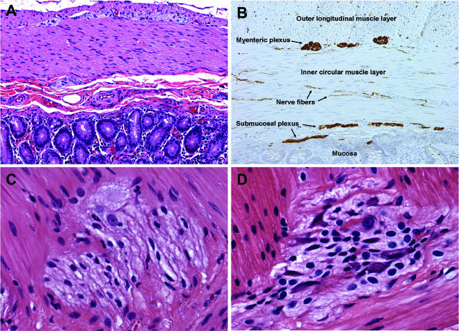
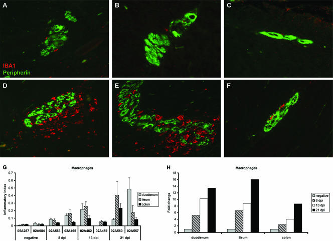
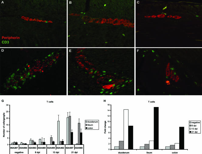
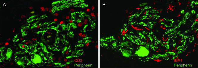
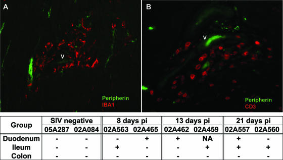
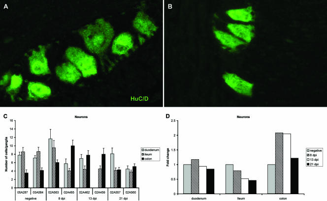
Gastrointestinal (GI) disease is a debilitating feature of human immunodeficiency virus (HIV) infection that can occur in the absence of histopathological abnormalities or identifiable enteropathogens. However, the mechanisms of GI dysfunction are poorly understood. The present study was undertaken to characterize changes in resident and inflammatory cells in the enteric nervous system (ENS) of macaques during the acute stage of simian immunodeficiency virus (SIV) infection to gain insight into potential pathogenic mechanisms of GI disease. Ganglia from duodenum, ileum, and colon were examined in healthy and acutely infected macaques by using a combination of routine histology, double-label immunofluorescence and in situ hybridization. Evaluation of tissues from infected macaques showed progressive infiltration of myenteric ganglia by CD3+ T cells and IBA1+ macrophages beginning as early as 8 days postinfection. Quantitative image analysis revealed that the severity of myenteric ganglionitis increased with time after SIV infection and, in general, was more severe in ganglia from the small intestine than in ganglia from the colon. Despite an abundance of inflammatory cells in myenteric ganglia during acute infection, the ENS was not a target for virus infection. This study provides evidence that the ENS may be playing a role in the pathogenesis of GI disease and enteropathy in HIV-infected people.
Figures
Similar articles
-
Expression of the simian Epstein-Barr virus-encoded latent membrane protein-1 in malignant lymphomas of SIV-infected rhesus macaques.J Med Virol. 2001 Sep;65(1):114-20. J Med Virol. 2001. PMID: 11505452
-
Simian immunodeficiency virus infection of the gastrointestinal tract of rhesus macaques. Functional, pathological, and morphological changes.Am J Pathol. 1993 Jun;142(6):1759-71. Am J Pathol. 1993. PMID: 8506946 Free PMC article.
-
Bone marrow monocyte/macrophages are an early cellular target of pathogenic and nonpathogenic isolates of simian immunodeficiency virus (SIVmac) in rhesus macaques.Lab Invest. 1995 Mar;72(3):323-33. Lab Invest. 1995. PMID: 7898051
-
Inflammatory neuropathies of the enteric nervous system.Gastroenterology. 2004 Jun;126(7):1872-83. doi: 10.1053/j.gastro.2004.02.024. Gastroenterology. 2004. PMID: 15188182 Review.
-
The role of prolonged viral gastrointestinal infections in the development of immunodeficiency-related enteropathy.Clin Rev Allergy Immunol. 2012 Feb;42(1):79-91. doi: 10.1007/s12016-011-8292-9. Clin Rev Allergy Immunol. 2012. PMID: 22116710 Review.
Cited by
-
Inflammation and epithelial cell injury in AIDS enteropathy: involvement of endoplasmic reticulum stress.FASEB J. 2011 Jul;25(7):2211-20. doi: 10.1096/fj.10-175992. Epub 2011 Mar 22. FASEB J. 2011. PMID: 21427211 Free PMC article.
-
Systemic gene delivery transduces the enteric nervous system of guinea pigs and cynomolgus macaques.Gene Ther. 2017 Oct;24(10):640-648. doi: 10.1038/gt.2017.72. Epub 2017 Aug 3. Gene Ther. 2017. PMID: 28771235 Free PMC article.
-
Sympathetic Nerves and Innate Immune System in the Spleen: Implications of Impairment in HIV-1 and Relevant Models.Cells. 2022 Feb 15;11(4):673. doi: 10.3390/cells11040673. Cells. 2022. PMID: 35203323 Free PMC article. Review.
-
Research Relevant Conditions and Pathology in Nonhuman Primates.ILAR J. 2020 Dec 31;61(2-3):139-166. doi: 10.1093/ilar/ilab017. ILAR J. 2020. PMID: 34129672 Free PMC article. Review.
-
Repeated occurrence of jejuno-jejunal intussusception in a calf.Can Vet J. 2009 Mar;50(3):287-90. Can Vet J. 2009. PMID: 19436481 Free PMC article.
References
-
- Barber, S. A., D. S. Herbst, B. T. Bullock, L. Gama, and J. E. Clements. 2004. Innate immune responses and control of acute simian immunodeficiency virus replication in the central nervous system. J. Neurovirol. 10(Suppl. 1):15-20. - PubMed
-
- Batman, P. A., A. R. Miller, P. M. Sedgwick, and G. E. Griffin. 1991. Autonomic denervation in jejunal mucosa of homosexual men infected with HIV. AIDS 5:1247-1252. - PubMed
-
- Bjarnason, I., D. R. Sharpstone, N. Francis, A. Marker, C. Taylor, M. Barrett, A. Macpherson, C. Baldwin, I. S. Menzies, R. C. Crane, T. Smith, A. Pozniak, and B. G. Gazzard. 1996. Intestinal inflammation, ileal structure and function in HIV. AIDS 10:1385-1391. - PubMed
-
- Boven, L. A., J. Middel, J. Verhoef, C. J. De Groot, and H. S. Nottet. 2000. Monocyte infiltration is highly associated with loss of the tight junction protein zonula occludens in HIV-1-associated dementia. Neuropathol. Appl. Neurobiol. 26:356-360. - PubMed
-
- Brookes, S. J. 2001. Classes of enteric nerve cells in the guinea-pig small intestine. Anat. Rec. 262:58-70. - PubMed
Publication types
MeSH terms
Substances
Grants and funding
LinkOut - more resources
Full Text Sources
