Comparative Study
. 2007 Jun;81(11):5949-57.
doi: 10.1128/JVI.00219-07. Epub 2007 Mar 28.

Structural basis for the recognition of blood group trisaccharides by norovirus

Affiliations
Comparative Study

Structural basis for the recognition of blood group trisaccharides by norovirus

Sheng Cao et al. J Virol. 2007 Jun.

Abstract

Noroviruses are one of the major causes of nonbacterial gastroenteritis epidemics in humans. Recent studies on norovirus receptors show that different noroviruses recognize different human histo-blood group antigens (HBGAs), and eight receptor binding patterns of noroviruses have been identified. The P domain of the norovirus capsids is directly involved in this recognition. To determine the precise locations and receptor binding modes of HBGA carbohydrates on the viral capsids, a recombinant P protein of a GII-4 strain norovirus, VA387, was cocrystallized with synthetic type A or B trisaccharides. Based on complex crystal structures observed at a 2.0-A resolution, we demonstrated that the receptor binding site lies at the outermost end of the P domain and forms an extensive hydrogen-bonding network with the saccharide ligand. The A and B trisaccharides display similar binding modes, and the common fucose ring plays a key role in this interaction. The extensive interface between the two protomers in a P dimer also plays a crucial role in the formation of the receptor binding interface.


Figures

FIG. 1.
FIG. 1.
Dimer of the VA387 P domain. (A) Ribbon representation of the P monomer and dimer. In the P monomer (left), the P1 subdomain is shown in green and the P2 subdomain is shown in red. The positions of Gly274 and Ala418 are marked with yellow spheres. In the P dimer (right), residue pairs (<3.3 Å apart) on the dimer interface are indicated by yellow stick models. (B) Topological diagram of the VA387 P domain. The arrows show the directions of β strands, whereas the α helix is represented by a cylinder. β strands in each β-sheet are colored identically. (C) Four layers of the dimer interface. One protomer is shown in a molecular surface model, and the other is in a light green, backbone trace model. Residues involved in hydrophobic, hydrophilic, and hydrogen-bonding interactions in the surface model are colored blue, red, and yellow, respectively. Three key secondary-structure elements on the dimer interface, β5, β9, and α1, are colored purple in the backbone model.
FIG. 2.
FIG. 2.
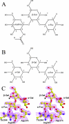
Trisaccharides from the complex crystal structures. (A) Chemical structure of the A trisaccharide. The asterisk denotes the position where the trisaccharide is connected to the rest of the HBGAs. Carbon atoms circling each saccharide ring are numbered. (B) Chemical structure of the B trisaccharide. The asterisk denotes the position where the trisaccharide is connected to the rest of the HBGAs. (C) Stereo view of the B trisaccharide. The B trisaccharide and three residues from the P domain in the vicinity of the α-Fuc ring are included. The superimposed (2FoFc) electron density map was calculated with the trisaccharide omitted from phasing and was contoured at 0.8σ with a 2.0-Å cover radius. The asterisks correspond to that in panel B.
FIG. 3.
FIG. 3.
α-Fuc binding site on the P domain dimer. (A and B) Side and top views of the antigen binding sites. Symmetry-related fucose rings are shown in sphere models together with the P dimer shown in the molecular surface model. (C) Surface cavity of the P dimer and the bound α-Fuc ring. The cavity interacting with the fucose ring is highlighted by the yellow ellipse, and the bottom of the cavity is circled in red. The orientation is similar to that in panel B. (D) Hydrogen-bonding network between the fucose ring and the P dimer. The fucose ring and residues involved in the interaction are shown in a ball-and-stick representation, with nitrogen, oxygen, and carbon atoms colored purple, red, and yellow, respectively. The dotted lines indicate hydrogen bonds. Backbones of the two protomers are shown as blue and green ribbons, respectively. The orientation is similar to that in panel A. Oδ1 and Oδ2, side chain groups of the Asp residue; Nε, Nɛ atom. (E) The hydrogen-bonding system for Arg345 and Asp374 on the protein side is shown in a stereo view. The orientation is similar to that in panel A.
FIG. 4.
FIG. 4.
Comparison of the P domains of VA387 and Norwalk virus. (A) Backbone superposition of the P protein of the VA387 strain (green) with that of Norwalk virus (purple; Protein Data Bank identification, 1IHM). The red regions represent sequence insertions (>3 amino acids) in the domain from the VA387 strain, and corresponding residue ranges are labeled along with the amino and carboxyl termini. (B) Structure-based sequence alignment of the P domain of VA387 and that of Norwalk virus. Boundaries between the hinge region, P1-1, P2, and P1-2 subdomains are marked as red triangles. Secondary-structure elements are shown for both structures, as assigned by the program DSSP (12). Identical and conserved residues are highlighted in blue and boxed with blue lines, respectively. Trisaccharide binding sites on the P protein are boxed with red lines. Regions missing in the final VA387 model are highlighted in yellow. Vertical arrows denote putative trypsin cleavage sites based on results from mass spectrometry analyses.
FIG. 5.
FIG. 5.
Alignment of sequences from strains representative of five binding patterns. Numbers denote the residue location in the primary sequence of VA387, where the key residues form the open binding cavity for α-Fuc. GenBank accession numbers are as follows: Snow Mountain virus (SMV), AAB61685; norovirus strain MOH, AAK84404; Hawaii virus, AAB97768; VA387, AAK84679; and VA207, AAK84676.

References

    1. Bertolotti-Ciarlet, A., S. E. Crawford, A. M. Hutson, and M. K. Estes. 2003. The 3′ end of Norwalk virus mRNA contains determinants that regulate the expression and stability of the viral capsid protein VP1: a novel function for the VP2 protein. J. Virol. 77:11603-11615. - PMC - PubMed
    1. Bertolotti-Ciarlet, A., L. J. White, R. Chen, B. V. Prasad, and M. K. Estes. 2002. Structural requirements for the assembly of Norwalk virus-like particles. J. Virol. 76:4044-4055. - PMC - PubMed
    1. Brunger, A. T., P. D. Adams, G. M. Clore, W. L. DeLano, P. Gros, R. W. Grosse-Kunstleve, J. S. Jiang, J. Kuszewski, M. Nilges, N. S. Pannu, R. J. Read, L. M. Rice, T. Simonson, and G. L. Warren. 1998. Crystallography and NMR system: a new software suite for macromolecular structure determination. Acta Crystallogr. D 54:905-921. - PubMed
    1. Chakravarty, S., A. M. Hutson, M. K. Estes, and B. V. Prasad. 2005. Evolutionary trace residues in noroviruses: importance in receptor binding, antigenicity, virion assembly, and strain diversity. J. Virol. 79:554-568. - PMC - PubMed
    1. Huang, P., T. Farkas, S. Marionneau, W. Zhong, N. Ruvoen-Clouet, A. L. Morrow, M. Altaye, L. K. Pickering, D. S. Newburg, J. LePendu, and X. Jiang. 2003. Noroviruses bind to human ABO, Lewis, and secretor histo-blood group antigens: identification of 4 distinct strain-specific patterns. J. Infect. Dis. 188:19-31. - PubMed

Publication types

MeSH terms