. 2007 Mar 28;27(13):3395-407.
doi: 10.1523/JNEUROSCI.4605-06.2007.

Robo1 and Robo2 cooperate to control the guidance of major axonal tracts in the mammalian forebrain

Affiliations

Robo1 and Robo2 cooperate to control the guidance of major axonal tracts in the mammalian forebrain

Guillermina López-Bendito et al. J Neurosci. .

Abstract

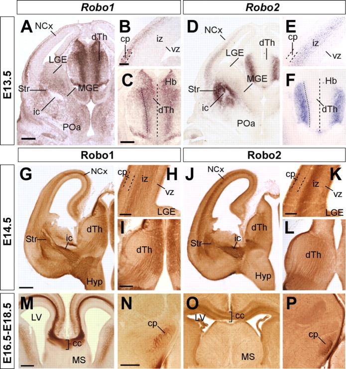
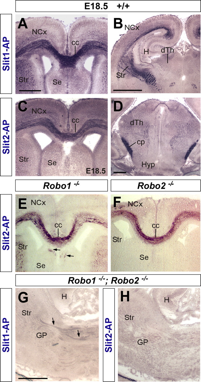
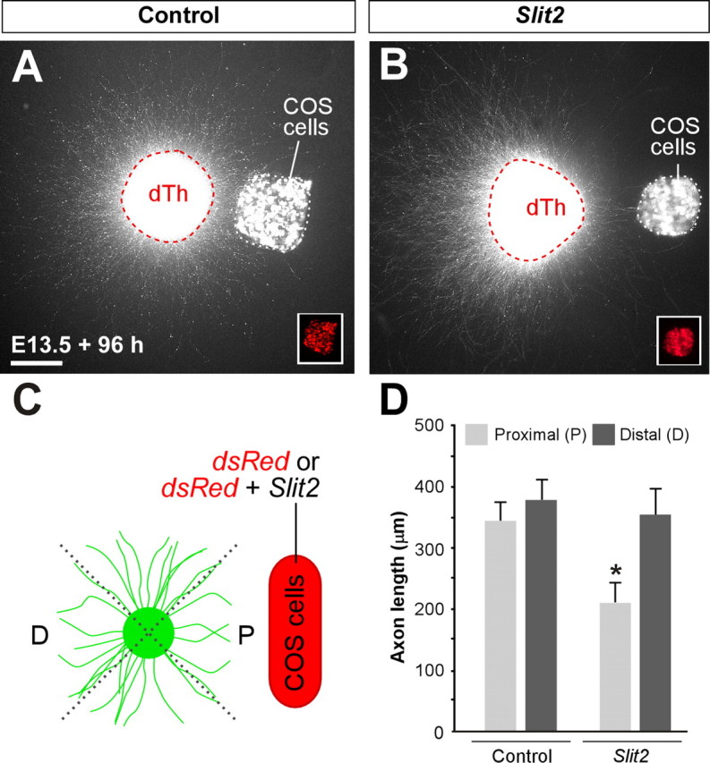
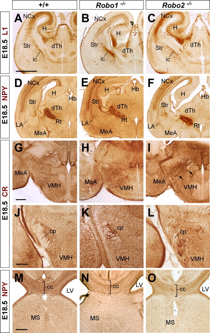
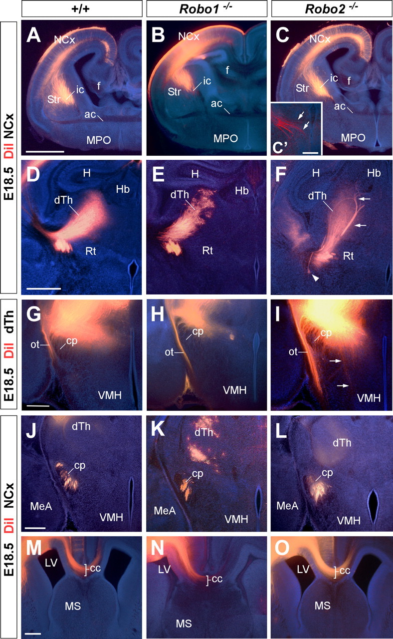
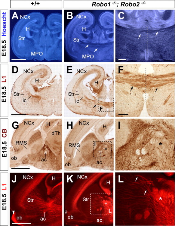
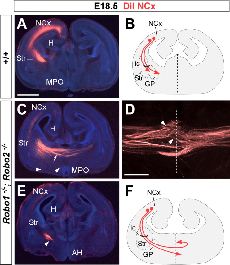
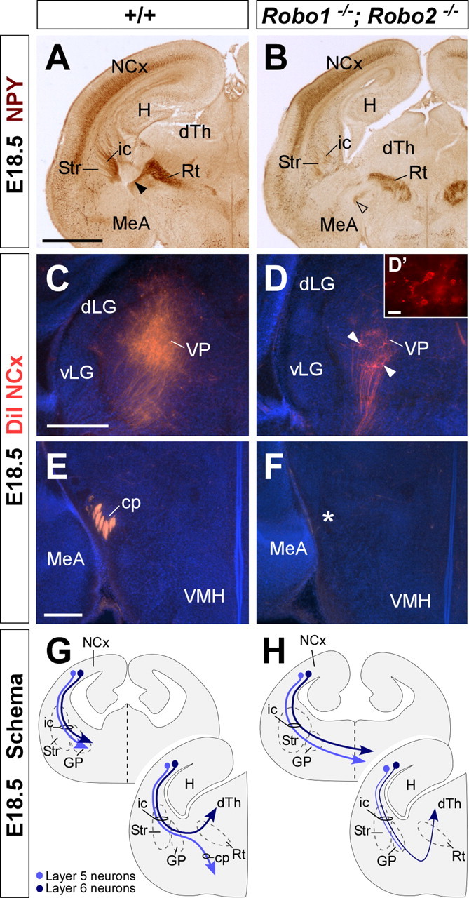
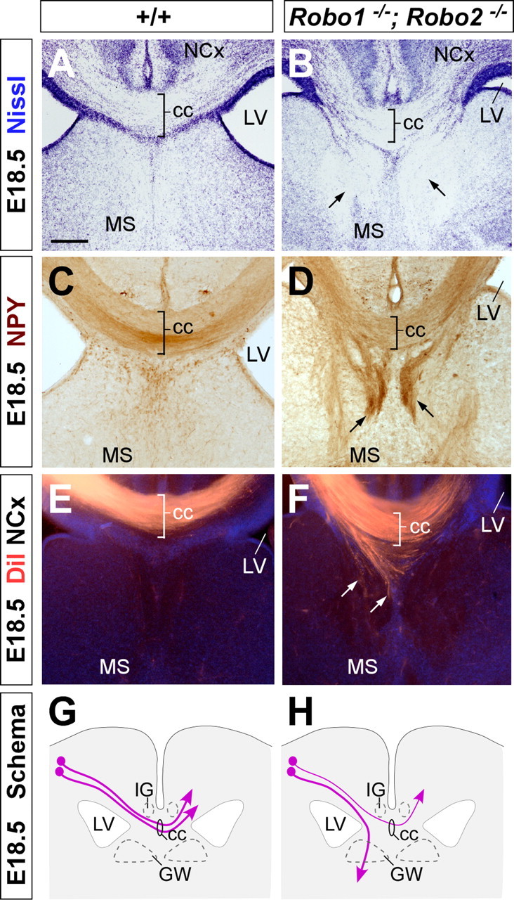
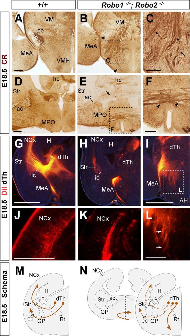
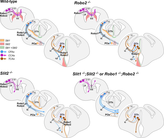
The function of the nervous system depends on the precision of axon wiring during development. Previous studies have demonstrated that Slits, a family of secreted chemorepellent proteins, are crucial for the proper development of several major forebrain tracts. Mice deficient in Slit2 or, even more so, in both Slit1 and Slit2 have defects in multiple axonal pathways, including corticofugal, thalamocortical, and callosal connections. In the spinal cord, members of the Robo family of proteins help mediate the function of Slits, but the relative contribution of these receptors to the guidance of forebrain projections remains to be determined. In the present study, we addressed the function of Robo1 and Robo2 in the guidance of forebrain projections by analyzing Robo1-, Robo2-, and Robo1;Robo2-deficient mice. Mice deficient in Robo2 and, more dramatically, in both Robo1 and Robo2, display prominent axon guidance errors in the development of corticofugal, thalamocortical, and corticocortical callosal connections. Our results demonstrate that Robo1 and Robo2 mostly cooperate to mediate the function of Slit proteins in guiding the major forebrain projections.


Figures

Figure 1.
Figure 1.
Expression of Robo1 and Robo2 receptors in the embryonic mouse forebrain. Serial coronal sections through mid-telencephalic/rostral diencephalic levels of an E13.5 embryo showing the expression of Robo1 and Robo2 mRNAs (A–D), and of E14.5, E16.5, and E18.5 embryos showing the expression of Robo1 (G–I, M, N) and Robo2 proteins (J–L, O, P). Robo1 and Robo2 mRNAs are expressed in the neocortex (NCx) and dorsal thalamus (dTh) in a partially complementary manner. Robo1 is expressed in the cortical plate (cp) (A, B), in a gradient decreasing from lateral to medial cortex, as well as in the dTh, in a gradient decreasing from the neuroepithelium to the mantle (A, C). Robo2 is expressed in the subplate and intermediate zone (iz) of the cortex (D, E), and in the most superficial region of the dTh (D, F). Coronal sections through E14.5, E16.5, and E18.5 brains showing Robo1 (G–I, M, N) and Robo2 (J–L, O, P) protein expression pattern are shown. Robo1 and Robo2 receptors are expressed in developing axons localized at the iz of the cortex (G, H, J, K), dorsal thalamus (I, L), and corpus callosum (cc) (M, O), and in the cerebral peduncle (cp) (N). ic, Internal capsule; Hb, habenula; vz, ventricular zone; Str, striatum; LGE, lateral ganglionic eminence; MGE, medial ganglionic eminence; MS, medial septum; LV, lateral ventricle; Hyp, hypothalamus; POa, anterior preoptic area. Scale bars: A, D, G, J, 300 μm; B, E, H, K, M, O, 100 μm; C, F, I, L, N, P, 200 μm.
Figure 2.
Figure 2.
Coronal sections through the forebrain of E18.5 wild-type (A–D), Robo1 (E), Robo2 (F), and Robo1;Robo2 double-mutant (G, H) fetuses showing Slit1–AP binding (A, B, G) and Slit2–AP binding (E, F, H). A–D, AP staining labels corticocortical axons at the corpus callosum (cc) (A, C), corticospinal axons at the cerebral peduncle (cp) (D), and thalamocortical/corticothalamic projections (B). E, F, Slit2–AP staining identifies corticocortical callosal projections in Robo1 (E) and Robo2 (F) mutant brains. Some ectopic bundles are abnormally displaced at the corpus callosum (cc) of Robo1 mutants (E, arrows). G, H, Slit–AP binding assays do not generally stain any axonal tract in the forebrain of Robo1;Robo2 double mutants. Only in a few cases, very weakly stained fibers could be observed after Slit1–AP binding (G, arrows). H, Hippocampus; Str, striatum; NCx, neocortex; SE, septum; dTh, dorsal thalamus; GP, globus pallidus; Hyp, hypothalamus. Scale bars: A, C, E, F, 500 μm; B, G, H, 1 mm; D, 200 μm.
Figure 3.
Figure 3.
Dorsal thalamic explants from E13.5 GFP transgenic mice showing Slit2 repulsion to dorsal thalamic axons. A, B, Dorsal thalamic explants were cocultured in collagen for 4 d in vitro either with mock-transfected COS cells (A) or Slit2-transfected COS cells (B). C, Scoring scheme used to test the effect of Slit2 on dorsal thalamus (dTh) axons in the experiments presented in A and B. Explants were subdivided into four equal sectors. The two sectors used for quantification were designated as proximal (P) and distal (D) in relation to the COS cell aggregate. The length of the 13 longest axons in each sector was measured in every explant. D, Quantification of the axonal growth of dTh explants cultured in collagen with mock-transfected COS cells (n = 19) or Slit2-transfected COS cells (n = 22) show a repulsive activity of Slit2. Mean length of axons was as follows: proximal, 345.8 ± 32.3 μm; distal, 374.9 ± 33.0 μm (average ± SEM) in controls; and proximal, 210.4 ± 29.4 μm; distal, 356.1 ± 43.5 μm (average ± SEM) in experimental cases. Significant differences were observed among proximal sectors in the case of Slit2 compared with controls cases. *p > 0.001. Scale bar, 200 μm.
Figure 4.
Figure 4.
Abnormal axonal trajectories in the forebrain of Robo1 and Robo2 single-mutant mice. Coronal sections through the telencephalon of E18.5 embryos showing cell adhesion molecule L1 (L1) (A–C), NPY (D–F, M–O), and calretinin (CR) (G–I) immunohistochemistry in wild-type (A, D, G, J, M), Robo1 (B, E, H, K, N), and Robo2 (C, F, I, L, O) mutant mice. A–C, In wild-type embryos, L1+ axons are confined to the intermediate zone of the neocortex (NCx), striatum (Str), and dorsal thalamus (dTh). In Robo1 and Robo2 mutants, L1+ fascicles are observed at the NCx and Str in a similar pattern as in wild-type embryos. D–F, Immunohistochemistry for NPY demonstrates that corticothalamic axons reach the diencephalon in wild-type (D), and Robo1 (E) and Robo2 mutant (F) brains. G–I, Coronal sections at the level of the diencephalon showing the trajectory of thalamocortical axons by immunohistochemistry for calretinin (CR) in wild-type (G), and Robo1 (H) and Robo2 (I) mutant brains. At this level, CR+ thalamocortical axons normally turn rostrally to enter the telencephalon, thus leaving the plane of section as observed in wild-type (G) and Robo1 mutants (H). In contrast, abnormal CR+ bundles were observed descending to the hypothalamus in Robo2 mutants (I, arrows). J–L, Abnormal development of the cerebral peduncle (cp) in Robo2 mutant brains (L), as revealed by calretinin immunostaining. M–O, Coronal sections at the level of the corpus callosum (cc) showing corticocortical fibers labeled by NPY immunohistochemistry in wild-type (M), and Robo1 (N) and Robo2 (O) mutant mice. ic, Internal capsule; H, hippocampus; Hb, habenula; Rt, reticular thalamic nucleus; VMH, ventromedial hypothalamic nucleus; MeA, medial amygdala; LA, lateral amygdala; MS, medial septum; LV, lateral ventricle. Scale bars: A–F, 1 mm; G–L, 100 μm; M–O, 200 μm.
Figure 5.
Figure 5.
Axon guidance defects in the forebrain of Robo2 single-mutant mice. Coronal sections through the telencephalon of E18.5 brains with DiI implanted in the neocortex (NCx) (A–F, J–O) or in the dorsal thalamus (dTh) (G–I) of wild-type (A, D, G, J, M), and Robo1 (B, E, H, K, N) and Robo2 (C, F, I, L, O) mutant mice, showing computer-generated overlays of DiI-labeled axons and Hoechst counterstain. A–C′, Coronal sections showing labeled DiI axons extending from the cortex into the internal capsule (ic) in wild-type (A), and Robo1 (B) and Robo2 (C, C′) mutant mice. D–F, Abnormal defasciculation (arrowhead) and targeting (arrows) of corticothalamic axons at the dorsal thalamus of Robo2 mutant mice (F). In Robo1 mutant mice, labeled axons extend from the cortex into the dorsal thalamus normally. G–I, Coronal sections through the caudal diencephalon showing dorsal thalamic fibers abnormally entering the hypothalamus in Robo2 mutant mice (I). No guidance defects were observed in the thalamocortical axons in Robo1 mutant mice (H) compared with wild type (G). J–L, Coronal sections showing corticospinal labeled axons at the cerebral peduncle (cp) of wild-type (J), and Robo1 (K) and Robo2 (L) mutant mice. Note the abnormal ventral position of the cerebral peduncle and the defasciculation of axons in Robo2 mutant mice. M–O, Rostral coronal sections showing corticocortical axons at the corpus callosum (cc) of wild-type (M), and Robo1 (N) and Robo2 (O) mutant mice. No guidance defects were observed in either of the Robo single mutants. ac, Anterior commissure; Str, striatum; f, fimbria; MPO, medial preoptic area; H, hippocampus; Rt, reticular thalamic nucleus; Hb, habenula; VMH, ventromedial hypothalamic nucleus; MeA, medial amygdala nucleus; MS, medial septum; ot, optic tract; LV, lateral ventricle. Scale bars: A–C, 1 mm; C′, 200 μm; D–F, 500 μm; G–O, 300 μm.
Figure 6.
Figure 6.
Abnormal axonal trajectories in the forebrain of Robo1;Robo2 double-mutant mice. Coronal (A–F) and sagittal (G–L) sections through the telencephalon of E18.5 embryos showing Hoechst staining (A–C), cell adhesion molecule L1 (D–F, J–L), and calbindin (G–I) immunohistochemistry in wild-type (A, D, G, J) and Robo1;Robo2 (B, C, E, F, H, I, K, L) mutant mice. A–C, Hoechst staining shows ectopic axonal bundles crossing the midline at the level of the medial preoptic region (MPO) (B, C, arrows). The anterior commissure (ac) is severely displaced dorsally. D–F, Immunostaining for L1 confirm that abnormal bundles of fibers cross the midline in Robo1;Robo2 mutants (E, F, arrows). G–I, Immunohistochemistry for calbindin delineates the abnormal crossing of the midline by unstained fibers (H, I). J–L, Ectopic bundles of axons crossing the midline are very evident in sagittal sections. Very few thalamic L1+ axons extend through the striatum in Robo1;Robo2 mutants because they accumulate in the midline (asterisk). The midline is indicated by a dotted line in C and F. NCx, Neocortex; ic, internal capsule; ob, olfactory bulb, RMS, rostral migratory steam; Str, striatum; H, hippocampus. Scale bars: A, B, D, E, G, H, J, K, 1 mm; C, 300 μm; F, I, L, 200 μm.
Figure 7.
Figure 7.
Corticofugal axons abnormally reach the telencephalic midline in Robo1;Robo2 double-mutant mice. Coronal sections through the telencephalon of E18.5 brains with DiI implanted in the neocortex (NCx), showing computer-generated overlays of DiI-labeled corticofugal axons and Hoechst counterstaining from wild-type (A) and Robo1;Robo2 mutants (C–E). The midline is indicated with a dotted line in D. The schemas summarize the results obtained in control (B) and Robo1;Robo2 mutants (F). A, B, In wild-type mice, labeled axons extend from the cortex into the striatum (Str). C–F, In Robo1;Robo2 mutants, labeled axons from the internal capsule (ic) abnormally approach the midline and cross it (C, arrow). A few axons that reach the midline course ventrally (arrowheads). Most of the axons that crossed the midline at more anterior levels were found in the contralateral side, where they either travel to the base of the telencephalon or extend toward the contralateral cortex (C, D). MPO, Medial preoptic area; H, hippocampus, AH, anterior hypothalamus. Scale bars: A, C, E, 1 mm; D, 300 μm.
Figure 8.
Figure 8.
Corticothalamic and corticospinal projections are severely defective in Robo1;Robo2 double-mutant mice. A, B, Coronal sections through the telencephalon of E18.5 brains showing NPY immunohistochemistry in wild-type (A) and Robo1;Robo2 (B) mutant mice. NPY immunohistochemistry demonstrates that some cortical axons reach the dorsal thalamus (dTh) in Robo1;Robo2 mutants, although through an abnormally ventral path (open arrowhead). C, D, Coronal sections through the dTh of E18.5 embryos with DiI implanted in the neocortex (NCx), showing computer-generated overlays of DiI retrogradely labeled cells and Hoechst counterstain from wild-type (C) and Robo1;Robo2 mutants (D). The number of retrogradely labeled cells found in the dTh is greatly reduced in Robo1;Robo2 mutants (D, arrowheads). E, F, The cerebral peduncle (cp) is absent in Robo1;Robo2 mutants (F), as revealed by the lack of DiI-labeled corticospinal fibers in the structure. The majority of these fibers cross abnormally the midline at more rostral levels. G, H, The schemas summarize the pathway followed by corticothalamic (layer 6; dark blue) and corticospinal (layer 5; light blue) axons in wild-type (G), and Robo1;Robo2 (H) mutant mice. Str, Striatum; Rt, reticular thalamic nucleus; VMH, ventromedial hypothalamic nucleus; MeA, medial amygdala; VP, ventroposterior nucleus; dLG, dorsal lateral geniculate nucleus, vLG, ventrolateral geniculate nucleus; ic, internal capsule; GP, globus pallidus; H, hippocampus. Scale bars: A, B, 1 mm; C, D, 300 μm; D′, 10 μm; E, F, 200 μm.
Figure 9.
Figure 9.
Abnormal development of the corpus callosum in Robo1;Robo2 double-mutant mice. Coronal sections through the telencephalon of E18.5 fetuses showing Nissl stain (A, B) and NPY (C, D) immunohistochemistry in wild-type (A, C) and Robo1;Robo2 double-mutant mice (B, D) mutant mice. A–D, Nissl staining and NPY immunohistochemistry demonstrates that Robo1;Robo2 double-mutant mice have a small corpus callosum (cc) and that large ectopic bundles of axons form on either side of it (B, D, arrows). E, F, Coronal sections through the telencephalon of E18.5 fetuses with DiI implanted in the neocortex (NCx), showing DiI-labeled corticocortical axons extending through the corpus callosum in wild-type (E) and Robo1;Robo2 double-mutant (F) mice. Note that many axons are abnormally directed ventrally before they reach the midline (F, arrows). G, H, The schemas summarize the pathways followed by corticocortical axons through the corpus callosum in wild-type (G) and Robo1;Robo2 double-mutant (H) mice. MS, Medial septum; LV, lateral ventricle; GW, glial wedge; IG, indusium griseum. Scale bars: A–F, 200 μm.
Figure 10.
Figure 10.
Thalamocortical axons follow abnormal paths in Robo1;Robo2 double-mutant mice. A–F, Coronal sections through the diencephalon of E18.5 fetuses showing calretinin immunohistochemistry in wild-type (A, D) and Robo1;Robo2 double-mutant mice (B, C, E, F). Note the abnormal invasion of thalamocortical fibers into the hypothalamus (C, arrows). At more rostral levels, thalamocortical axons cross the ventral midline (F, arrowheads) below the additional abnormal cross of corticofugal fibers (E, arrow). G–I, L, Coronal sections through the diencephalon of E18.5 embryos with DiI implanted in the dorsal thalamus (dTh), showing DiI axons abnormally entering the hypothalamus in Robo1;Robo2 double-mutant mice (H, I). J, K, Very few thalamic axons reach the cortex in Robo1;Robo2 double-mutant mice. M, N, The schemas summarize the pathways followed by thalamocortical axons in wild-type (M) and Robo1;Robo2 double-mutant mice (N). Str, Striatum; VMH, ventromedial hypothalamic nucleus; MeA, medial amygdala; cp, cerebral peduncle; ac, anterior commissure; ic, internal capsule; ec, external capsule; GP, globus pallidus; Rt, reticular thalamic nucleus; MPO, medial preoptic area; hc, hippocampal commissure; H, hippocampus, AH, anterior hypothalamus; VM, ventromedial nucleus. Scale bars: A, B, D, E, 300 μm; C, F, 200 μm; G–I, 1 mm; J–L, 200 μm.
Figure 11.
Figure 11.
Schematic summarizing axonal defects in the Slit and Robo mutants in relation to regions of Slit action and Robo expression. Slit2 acts in ventral and ventrolateral domains, and Slit1 action is limited to medial areas. In wild-type animals, Slit2 prevents Robo-expressing axons from entering ventral regions and also prevents the axons from coursing medially. Loss of Slit1 does not have an impact on the axonal trajectories because of the continued presence of Slit2. However, loss of Slit2 allows the axons to project ventrally and medially, potentially in response to positive cues from these areas. Mice lacking Robo2 have a less severe phenotype than the one observed in Slit2−/− because of the presence of Robo1 in those axonal tracts. Only a few corticofugal axons are misrouted from their normal route at the ventral telencephalon. Loss of Robo1 and Robo2 mimics the phenotype observed in mice lacking both Slit1 and Slit2, and allows axons to travel ventrally and medially and to approach and cross the midline. LV, Lateral ventricle; cc, corpus callosum; DTh, dorsal thalamus; ic, internal capsule; cp, cortical plate; POa, anterior preoptic area; II–VI, cortical layers.

References

    1. Andrews W, Liapi A, Plachez C, Camurri L, Zhang J, Mori S, Murakami F, Parnavelas JG, Sundaresan V, Richards LJ. Robo1 regulates the development of major axon tracts and interneuron migration in the forebrain. Development. 2006;133:2243–2252. - PubMed
    1. Auladell C, Pérez-Sust P, Supèr H, Soriano E. The early development of thalamocortical and corticothalamic projections in the mouse. Anat Embryol. 2000;201:169–179. - PubMed
    1. Bagri A, Marín O, Plump AS, Mak J, Pleasure SJ, Rubenstein JL, Tessier-Lavigne M. Slit proteins prevent midline crossing and determine the dorsoventral position of major axonal pathways in the mammalian forebrain. Neuron. 2002;33:233–248. - PubMed
    1. Braisted JE, Tuttle R, O'Leary DD. Thalamocortical axons are influenced by chemorepellent and chemoattractant activities localized to decision points along their path. Dev Biol. 1999;208:430–440. - PubMed
    1. Braisted JE, Catalano SM, Stimac R, Kennedy TE, Tessier-Lavigne M, Shatz CJ, O'Leary DD. Netrin-1 promotes thalamic axon growth and is required for proper development of the thalamocortical projection. J Neurosci. 2000;20:5792–5801. - PMC - PubMed

Publication types

Substances